


Traditional pacing modes
Depending on the number of leads and the device model, different pacing modes can be programmed. Choosing the mode is the first essential step in programming a single- or dual-chamber pacemaker, since it directly affects the other parameters; it takes into account the patient’s age, indication for implantation, intrinsic rhythm, exercise capacity, chronotropic response and comorbidities. The clinician in charge of follow-up must know how each mode works, with its advantages and limitations.
Content
The international NBG code
Combined efforts by the North American (NASPE) and British (BPEG) societies of pacing and electrophysiology have defined the international NBG code (last revised in 2002). Each mode is designated by a 4- or 5-letter code describing its basic operation; in practice the code is shortened to 3 or 4 letters, and most pacemakers are programmed in VVI, VVIR, DDD or DDDR.
| Letter | Describes | Codes |
|---|---|---|
| 1st | Chamber(s) paced | V = ventricle, A = atrium, D = dual (A + V), O = none |
| 2nd | Chamber(s) sensed | V, A, D; O = none (asynchronous operation) |
| 3rd | Response to sensing | I = inhibited, T = triggered, D = dual (I + T), O = none |
| 4th | Rate modulation | R = rate response (sensor); simply omitted when off: DDD, not DDDO |
| 5th (optional, rarely used) | Multisite pacing | O = none, A = atrium, V = ventricle, D = dual (A + V) |
- Inhibited (I): a sensed intrinsic event withholds pacing.
- Triggered (T): a sensed event triggers pacing (AAT, VVT). In the dual (D) response of VDD and DDD, an atrial sensed event triggers ventricular pacing if no ventricular event is sensed within the AV delay (atrial tracking).
- DDI is a non-tracking mode: an atrial sensed event never triggers ventricular pacing.
- O is the letter O, not the digit zero: AOO, VOO, DOO, ODO.
- S (single chamber, A or V) is a manufacturers’ designation only: SSI = AAI or VVI, depending on the lead position.
Single-chamber pacing
Asynchronous modes: AOO and VOO
- Single-chamber pacing
- No sensing
- Asynchronous pacing
AOO and VOO deliver asynchronous pacing at the programmed lower rate: with no sensing, intrinsic events never inhibit pacing (regular spikes on the ECG), and the only timing parameter is the pacing rate (rate = 60,000 / pacing interval in ms). Asynchronous modes (AOO, VOO, DOO) are the most primitive pacing modes, the only ones available in the first programmable devices.
In non-dependent patients, spontaneous and paced activity compete (parasystole) and stimuli may fall in the vulnerable period, with a risk of atrial fibrillation (atrial pacing) or, very rarely outside myocardial ischemia or metabolic disorders, ventricular fibrillation (R-on-T). A rate above the spontaneous rate promotes capture and reduces, without abolishing, this risk. Asynchronous modes are therefore used only temporarily: in pacing-dependent patients undergoing surgery with electrocautery (to avoid inhibition and asystole) or an MRI scan, and as the classical magnet response (see below).
VVI/VVIR mode
- Ventricular pacing
- Ventricular sensing
- A ventricular event sensed outside the refractory period inhibits pacing
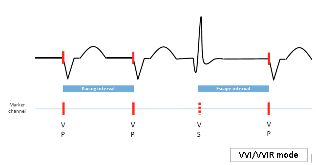
VVI mode provides single-chamber ventricular pacing at the programmed lower rate unless inhibited by a sensed ventricular event, each sensed or paced event starting a new escape interval; in VVIR mode, the rate follows the patient’s activity measured by a sensor. Ventricular sensing often occurs after the onset of the surface QRS, especially with right bundle branch block (the signal is sensed only when activation reaches the right ventricular lead). Signals falling in the refractory period that follows each sensed or paced event do not reset the escape interval, which prevents double sensing of the QRS and T-wave sensing.
Schematic example of VVI
Clinical example of VVI
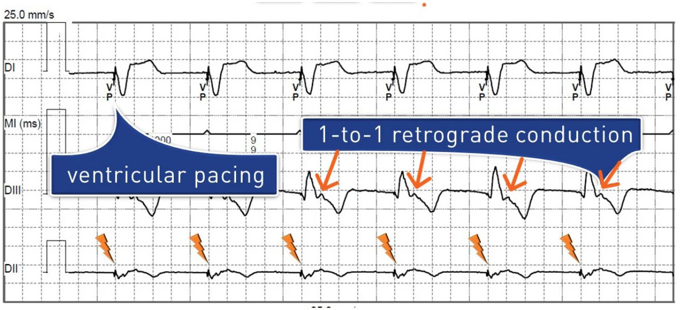
VVI prevents ventricular bradycardia and is primarily indicated in atrial fibrillation with a slow ventricular response, where atrial sensing and pacing are useless. With a lower rate below the spontaneous rate (e.g., 40 bpm), it works in sentinel mode, pacing only when required, which suits patients needing infrequent pacing. In sinus node dysfunction, a lower rate above the sinus rate makes ventricular pacing permanent, with retrograde conduction when VA conduction is intact: the atria contract against closed AV valves, with retrograde flow into the pulmonary veins and venae cavae, which can cause pacemaker syndrome (dyspnea, orthopnea, neck pulsations, palpitations, chest pain).
AAI/AAIR mode
- Atrial pacing
- Atrial sensing
- An atrial event sensed outside the refractory period inhibits pacing
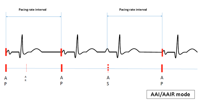
AAI, the atrial counterpart of VVI, provides atrial pacing at the lower rate unless inhibited by an atrial sensed event (sensor rate in AAIR). Programming is nearly the same as in VVI, but atrial signals are smaller, requiring a higher sensitivity (lower programmed value), and the refractory period must be longer to prevent far-field R-wave oversensing, which would slow the pacing rate by resetting the escape interval. Such oversensing is corrected by reducing atrial sensitivity and/or lengthening the refractory period.
Schematic example of AAI
Clinical example of AAI
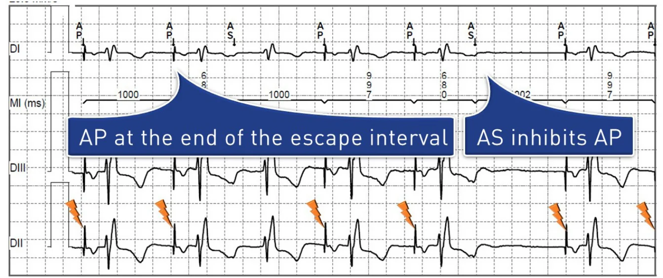
The preferred indication of AAI(R) is pure sinus node dysfunction. It is formally contraindicated in permanent or paroxysmal AV conduction disorders and should be avoided in patients with vagal syncope or carotid sinus syndrome. If it is considered, AV conduction must be assessed: 1:1 AV conduction during atrial pacing at 120–130 bpm, with a normal PR interval and normal QRS complexes, is a good sign of preserved conduction.
Triggered modes: AAT and VVT
- Atrial or ventricular pacing
- Atrial or ventricular sensing
- A sensed event outside the refractory period triggers immediate pacing in the same chamber
In AAT and VVT, pacing occurs at the lower rate, but a sensed event outside the refractory period triggers an immediate pulse in the same chamber instead of inhibiting it; this stimulus is harmless, falling in the natural refractory period of the myocardium. The maximum pacing rate and the refractory period prevent the device from running away with rapid signals. On the ECG in VVT, a spike appears within each sensed QRS, whose morphology is unchanged (pseudo-fusion) or slightly altered (fusion); AAT is the atrial equivalent.
Useful in older pacemakers with unipolar sensing, highly susceptible to interference, triggered modes are now rarely used as a primary mode (they also waste battery energy). They remain useful temporarily to prevent inhibition in a pacemaker-dependent patient during surgery with electrocautery, and for diagnosis: each spike marks the moment of sensing on the ECG, to check sensing and the absence of far-field oversensing. The dual-chamber triggered mode, DDT, is likewise a temporary sensing test mode.
Dual-chamber pacing
ODO: sensing without pacing
In ODO mode, the device senses the atrium and the ventricle but never paces, whatever the intrinsic rate; markers and electrograms show sensing in both chambers (in the example patient: sinus rhythm sensed AS-VS, wide QRS complexes and premature atrial contractions). ODO is programmed only temporarily, for diagnostic purposes such as checking sensing, or in non-pacing-dependent patients scheduled for an MRI scan; never permanently, for safety reasons, unless pacing is considered detrimental.
DOO: asynchronous dual-chamber pacing
- Asynchronous dual-chamber pacing
- No sensing
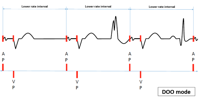
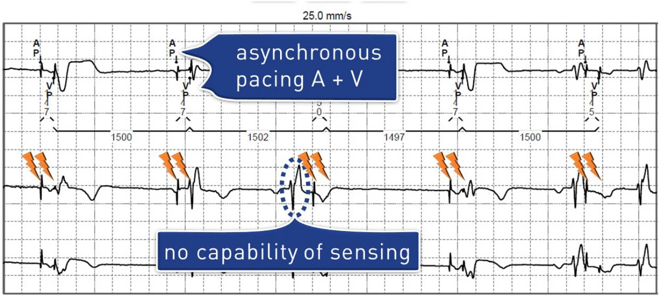
DOO mode delivers asynchronous AV sequential pacing at the programmed lower rate, without inhibition by intrinsic atrial or ventricular events. The AV and VA intervals are fixed and are not reset, since sensing is not possible. Its risks and uses are those of the asynchronous modes described above.
Schematic example of DOO
Clinical example of DOO
VDD mode
- Ventricular pacing
- Dual-chamber sensing (atrium and ventricle)
- Atrial sensing triggers ventricular pacing after the AV delay; ventricular sensing inhibits it
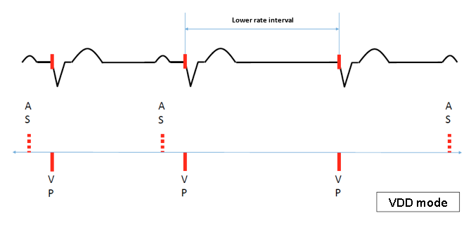
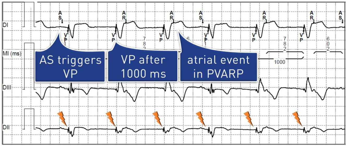
VDD senses the atrium and the ventricle but paces only the ventricle. It is a tracking mode: an atrial sensed event triggers ventricular pacing after the AV delay, up to the maximum tracking rate. When the atrial rate is slower than the lower rate, VVI pacing is delivered at the lower rate, since atrial pacing is impossible. VDD is mostly used with single-lead systems, a ventricular lead sensing the atrium through two floating electrodes on its intra-atrial portion, but it is also a programmable option in many dual-chamber pacemakers.
Schematic example of VDD
Clinical example of VDD
VDD may be indicated in complete AV block with preserved sinus node function, not in sinus node dysfunction. A low lower rate, possibly with rate hysteresis, promotes spontaneous atrial activity and avoids VVI behavior and retrograde conduction. With a floating dipole, atrial sensing is sometimes less reliable than with an atrial lead, especially during deep breathing and exercise, which also degrades atrial arrhythmia detection and memory data; in AV block, loss of atrial sensing on exertion causes a sudden drop to the lower rate, with possible pacemaker syndrome and a risk of pacemaker-mediated tachycardia. VDD is now also available in some leadless pacemakers, whose accelerometer detects atrial contraction.
DDI/DDIR mode
- Dual-chamber pacing (atrium and ventricle)
- Dual-chamber sensing (atrium and ventricle)
- Sensed events inhibit pacing (atrial and ventricular); no atrial tracking
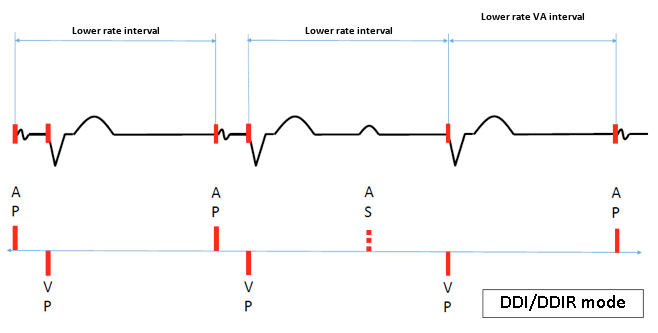
DDI can be thought of as DDD without P-wave tracking. Atrial pacing occurs at the lower rate, followed by an AV delay and ventricular pacing if no ventricular event is sensed. A sensed atrial event inhibits atrial pacing but, unlike in DDD, starts no AV delay: AV synchrony is kept with atrial pacing but lost in AV block with sensed P waves, the ventricle then being paced at the lower rate, dissociated from atrial activity (pseudo-VVI behavior). The paced rate never exceeds the lower rate (or the sensor rate in DDIR), so fast atrial arrhythmias never cause rapid ventricular pacing.
Schematic example of DDI
Clinical example of DDI
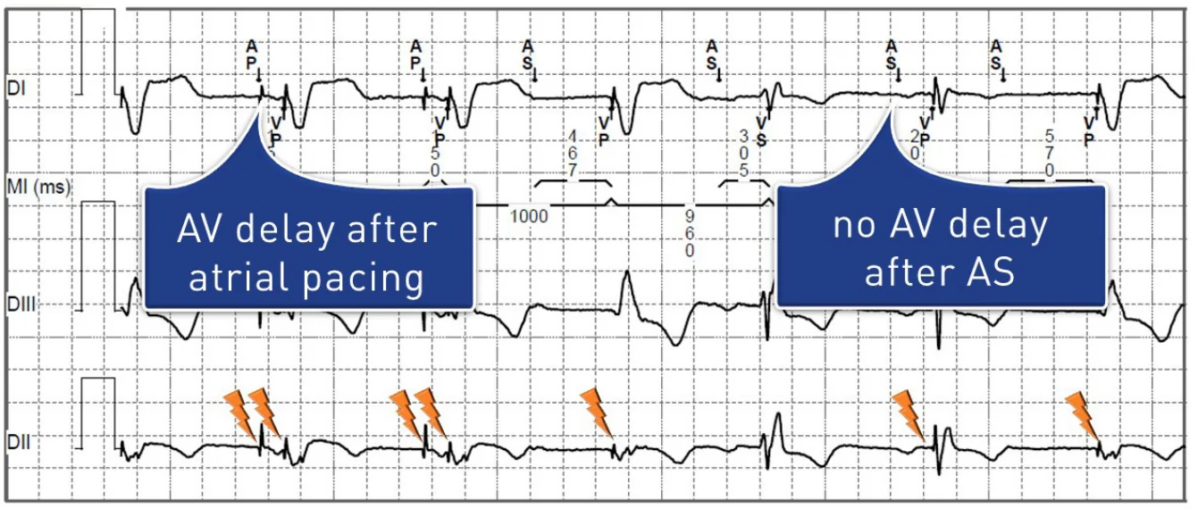
The lower rate should be high enough, possibly with rate response, to favor atrial pacing and avoid spontaneous P waves, pseudo-VVI behavior and retrograde conduction. DDI is commonly used as the mode-switch mode, to avoid rapid ventricular pacing during atrial arrhythmias. It can be programmed long term in patients combining complete AV block, fast AF episodes and sinus node dysfunction (AP-VP pacing), is useful when mode switching works suboptimally, and may be considered in patients with repeated pacemaker-mediated tachycardia. It does not suit complete AV block with a normal, faster sinus rate, since P waves start no AV delay.
DDD/DDDR mode
- Dual-chamber pacing (atrium and ventricle)
- Dual-chamber sensing (atrium and ventricle)
- Dual response to sensing (inhibition and triggering)
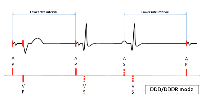
DDD is the most commonly used pacing mode, appropriate for sinus node dysfunction and/or AV block, and the standard mode of dual-chamber and resynchronization pacemakers; it maintains AV synchrony at rest and during exercise. Intrinsic atrial or ventricular activity outside the refractory periods inhibits pacing in that chamber, and each paced atrial event, or atrial event sensed outside the refractory periods, starts an AV delay ending with ventricular pacing if no ventricular event is sensed. DDD can thus deliver AP-VP, AP-VS, AS-VP (atrial tracking) or AS-VS (complete inhibition) sequences.
AV delays after sensed and paced atrial events are programmed separately; they can shorten at higher rates (rate-adaptive AV delay) or lengthen to favor intrinsic conduction. Ventricular pacing tracks sensed atrial events up to the maximum tracking rate, above which the ventricle is no longer paced at the atrial rate. In DDDR mode, the pacemaker follows the faster of the atrial and sensor rates; the maximum tracking rate and the maximum sensor rate are programmed separately.
Schematic example of DDD
Clinical example of DDD
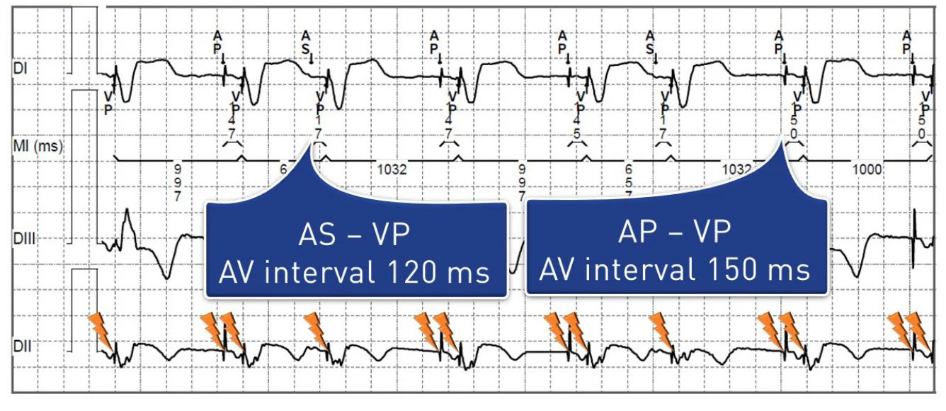
In this patient, follow-up would probably show a percentage of ventricular pacing close to 100%. One of the priorities of programming is to avoid unnecessary right ventricular pacing without compromising patient safety.
Avoiding unnecessary right ventricular pacing
Prolonged right ventricular pacing promotes deleterious remodeling (increased ventricular volumes) and atrial arrhythmias, and reducing it prolongs battery life. A high percentage of ventricular pacing in a patient implanted for sinus node dysfunction should therefore prompt a discussion of specific algorithms. Permanently long AV delays reduce ventricular pacing but impair ventricular filling and favor pacemaker-mediated tachycardia and 2:1 block on exercise. Other options are AV hysteresis (e.g., VIP, Abbott) and modes switching between AAI/ADI and DDD: MVP (Medtronic), RYTHMIQ (Boston Scientific), SafeR (MicroPort) and Vp Suppression (Biotronik). With these modes, occasional blocked P waves can be normal operation; they should be avoided in high-grade AV block.
Overview of pacing modes
| Mode | Key use | Main risks / limits |
|---|---|---|
| ODO | Diagnostics (sensing check); MRI in non-pacing-dependent patients | No pacing: never for long-term use |
| AOO / VOO / DOO | Surgery with electrocautery or MRI in pacing-dependent patients; magnet mode | Pacing in the vulnerable period (R-on-T, AF) |
| AAT / VVT | Older devices; surgery; sensing checks | Rarely used today |
| AAI(R) | Pure sinus node dysfunction | Contraindicated in AV conduction disorders; DDD(R) preferred (ESC 2021) |
| VVI(R) | AF with slow ventricular response | Pacemaker syndrome (retrograde conduction) |
| VDD | AV block with preserved sinus node function | Less reliable atrial sensing (floating dipole) |
| DDI(R) | Atrial arrhythmias (mode switch); repeated pacemaker-mediated tachycardia | No AV synchrony on sensed P waves |
| DDD(R) | Sinus node dysfunction and/or AV block | Unnecessary right ventricular pacing |