


Algorithms favoring AV conduction
Content
Why avoid unnecessary right ventricular pacing?
Extensive literature demonstrates the deleterious effect of prolonged right ventricular pacing on left ventricular function: it induces deleterious ventricular remodeling, with increased ventricular volumes and an increased risk of atrial arrhythmia. Similarly, any reduction in unnecessary ventricular pacing saves battery, prolongs the life of the device and may reduce the frequency of battery replacement. Manufacturers have proposed various ways of reducing the percentage of ventricular pacing without inducing symptoms or compromising patient safety in the event of an atrioventricular conduction disorder. The preferred indication for this type of mode is therefore sinus node dysfunction with preserved or paroxysmally altered atrioventricular conduction.
- Evaluation of the ventricular pacing burden is an important component of follow-up in a patient with sinus node dysfunction, with the aim of reducing to a minimum any unnecessary ventricular pacing.
- A high percentage of right ventricular pacing in a patient implanted for sinus node dysfunction with preserved AV conduction should alert the physician at interrogation and prompt a discussion of specific algorithms.
- These modes should be avoided in patients with high-grade AV block.
Three ways to reduce ventricular pacing
- Permanent long AV delays may prevent ventricular pacing, but may also sacrifice optimal end-diastolic preload by the atria, increase the risk of pacemaker-mediated tachycardia (PMT) and favor 2:1 block during exercise.
- AV delay hysteresis intermittently prolongs the AV delay to promote intrinsic activation. If an R wave is sensed, the AV delay remains prolonged; if not, the ventricle is paced at the end of the extended delay (no blocked atrial activity) and the programmed AV delay resumes. Older than the dedicated modes, it is still available from all manufacturers; this is how Abbott’s VIP works.
- Dedicated pacing modes switch between AAI/ADI and DDD: MVP (Medtronic), RYTHMIQ (Boston Scientific), SafeR (MicroPort) and Vp-Suppression (Biotronik). They are easy to program; their general philosophy is the same, but their operating details differ and must be perfectly understood.
The common principle: AAI/ADI pacing with a switch to DDD
In these dedicated modes, the device paces in AAI(R) mode while analysing ventricular sensing (ADI mode). All of them provide:
- AAI pacing (ADI mode) as long as AV conduction is preserved;
- an automatic switch to DDD(R) pacing when AV conduction is altered, with manufacturer-specific criteria (blocked atrial events, slow ventricular cycles, long PR interval or ventricular pause); MVP and RYTHMIQ also deliver back-up ventricular pacing before switching;
- periodic searches for intrinsic conduction while in DDD mode, with automatic return to AAI/ADI when AV conduction resumes.
Classic pitfalls
Blocked P waves and ventricular pauses
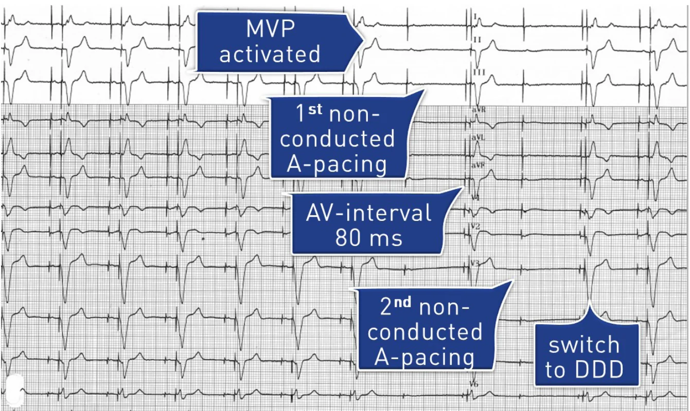
When AV conduction fails in AAI/ADI operation, the blocked atrial event is followed by a ventricular pause until back-up pacing or the switch to DDD. With the first version of MVP, the maximal pause is about twice the lower rate interval (approximately 2 seconds at 60 bpm, 4 seconds at 30 bpm). This pause may be symptomatic and can favor pause-dependent ventricular arrhythmias (long–short sequence), so a lower rate limit of 50 bpm or higher is recommended. To shorten the pause, the updated MVP paces the atrium 420 to 600 ms after the blocked atrial activity, then the ventricle 80 ms later.
Long PR interval
A long PR interval with 1:1 conduction produces no blocked P wave, so the first version of MVP never switched to DDD, even in patients with symptoms on exertion. The updated MVP, SafeR and Vp-Suppression can switch to DDD on a long PR/AR interval (see table); in MVP this function is OFF by default and probably to be reserved for patients with symptoms on exertion. Permanent DDD(R) may be more appropriate for symptomatic first-degree AV block, particularly if the PR interval does not shorten during exercise.
PMT and 2:1 block during AV-delay searches
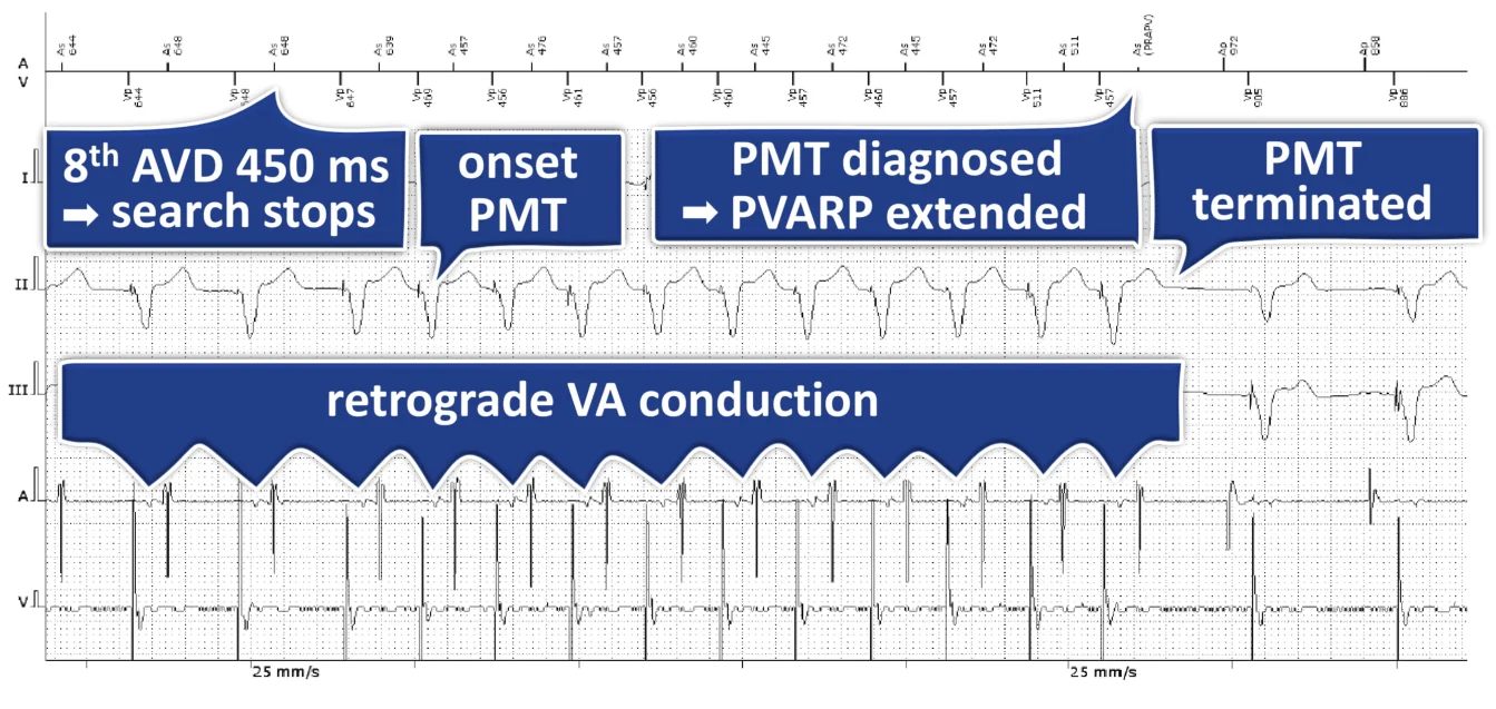
When the search for intrinsic conduction relies on a prolonged AV delay (VIP, AV Search+, Vp-Suppression), no P wave can be blocked, because the ventricle is paced at the end of the extended AV delay. However, the prolonged AV delay favors retrograde conduction and PMT, and lengthens the total atrial refractory period (AV delay + PVARP), so 2:1 block may occur during exercise; by contrast, the risk of PMT is nil in AAI mode with VVI back-up. In patients with impaired anterograde but preserved retrograde conduction, it is advisable not to program these searches, or at least to verify that the PVARP is longer than the retrograde conduction time; in permanent complete AV block, the simplest approach is not to program the algorithm. A long AV delay also favors repetitive non-reentrant ventriculoatrial synchrony (RNRVAS).
Premature beats and sensing: inappropriate switches
Inappropriate switches to DDD are relatively common and increase the percentage of ventricular pacing. Frequent causes are the compensatory pause after a PVC, dropped premature atrial contractions or atrial runs, late PVCs falling in the post-atrial pacing ventricular blanking or noise window, and an active junctional rhythm mimicking a long PR interval. Correct sensing and effective atrial pacing are prerequisites: ventricular undersensing leads to back-up pacing and an inappropriate switch. MicroPort and Boston Scientific devices record an EGM at the switch, which allows the diagnosis to be verified.
The five algorithms compared
| Algorithm (company) | How it works | Switch to DDD | Return to intrinsic conduction | Main pitfall |
|---|---|---|---|---|
| MVP (Medtronic) | AAI(R) ↔ DDD(R): AAI pacing while monitoring AV conduction (ADI). A single blocked P wave is tolerated (never two in a row); on the next cycle, back-up ventricular pacing 80 ms after the scheduled atrial pace (signature). | No ventricular sensed event in 2 of the 4 preceding A-A intervals. Updated version: also when the average PR/AR interval exceeds a programmable limit (OFF by default). | Check (one AAI cycle) 1 min after the switch; after each failure the interval doubles (2, 4, 8… min, up to 16 h). | First version: pause up to about twice the lower rate interval, and no switch on long PR. Program a lower rate ≥ 50 bpm. |
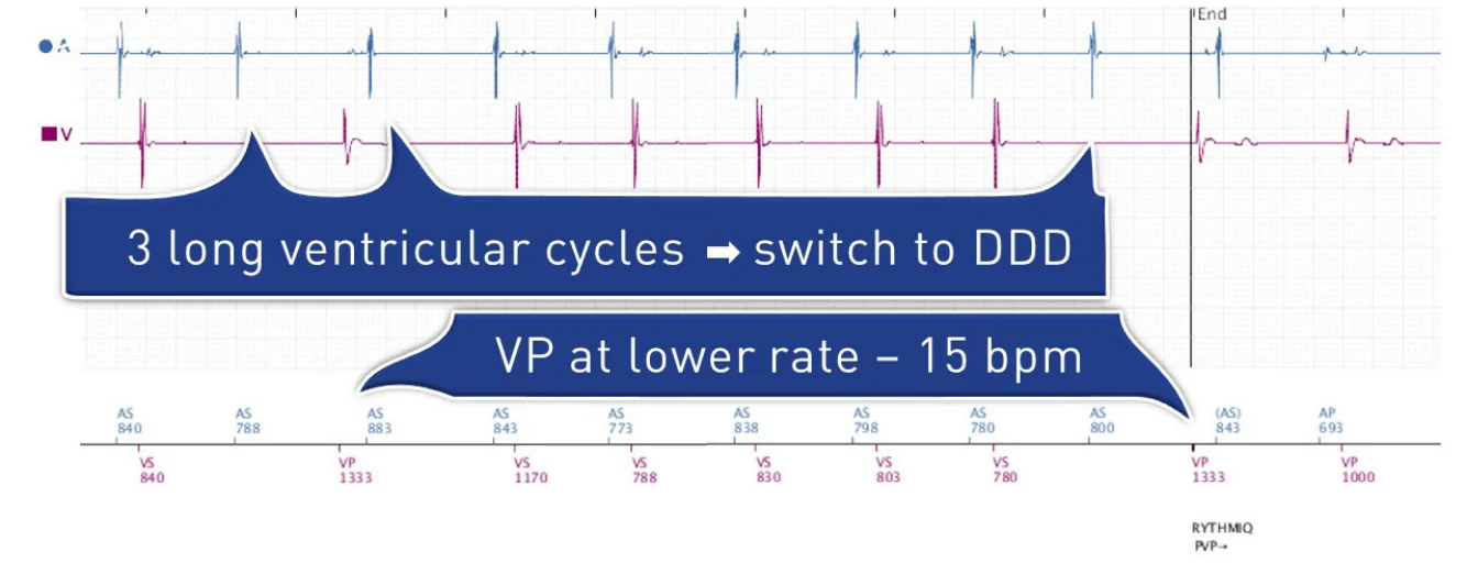
| RYTHMIQ (Boston Scientific) | AAI(R) with VVI back-up pacing 15 bpm below the lower rate limit (within 30–60 bpm). Nominally OFF. | 3 slow ventricular beats in a rolling window of 11 (a paced beat, or a sensed beat more than 150 ms longer than the lower rate or sensor interval); blocked P waves are not counted. | AV Search+ (AV delay hysteresis every 32–1024 cycles): back to AAI(R) with VVI back-up after ≥ 25 VS-Hy beats. If AV Search+ is OFF, the device stays in DDD(R). | Inappropriate switches with PVCs/PACs and late PVCs in the post-atrial blanking or noise window; PMT and 2:1 block during AV Search+. |
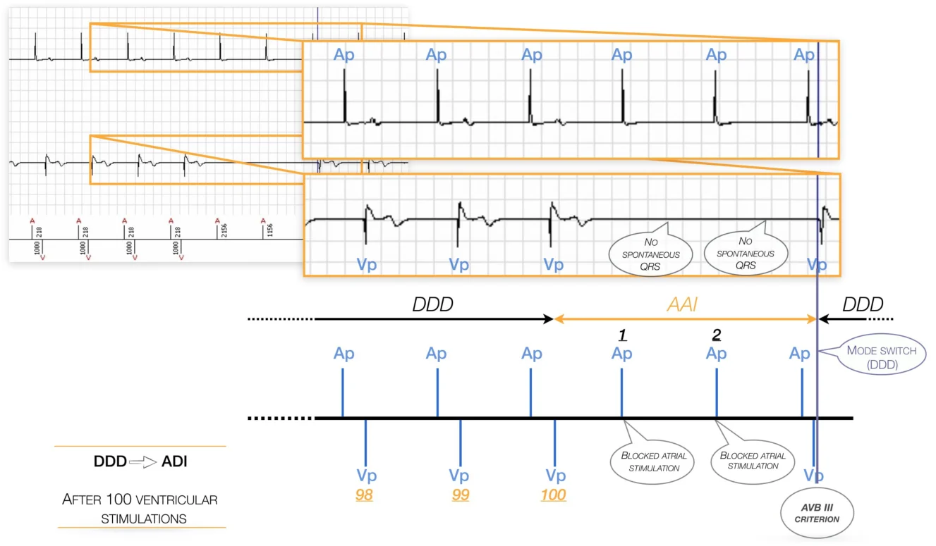
| SafeR (MicroPort) | ADI mode; one switch criterion for each degree of AV block, plus a pause criterion. | AVB III: 2 consecutive blocked atrial events; AVB II: 3 non-consecutive in the last 12 cycles; AVB I: PR/AR above the programmed limit for 6 consecutive cycles; pause > 2, 3 or 4 s. | After 12 consecutive spontaneous ventricular events, or automatically every 100 paced ventricular cycles; if AV block is frequent, stays in DDD until 8:00 am the next morning. | Blocked P waves at each return to ADI; false AVB I (junctional rhythm), false AVB II (PACs, atrial runs) or false pause (PVCs, especially with the 2 s setting). |
| VIP (Abbott) | No specific mode: AV delay hysteresis. The AV delay is extended by 50–200 ms (total ≤ 450 ms) for 1–3 cycles, every 30 s to 30 min; the ventricle is paced at the end of the extended delay, so no P wave is blocked. | None (DDD throughout); the programmed AV delay resumes after the programmed number of consecutive paced cycles. Suspended at rates ≥ 110 bpm. | VIP is activated when an R wave is sensed during a search, or after 3 consecutive sensed R waves outside a search. | Increased PMT risk: avoid when anterograde conduction is impaired but retrograde conduction preserved. |
| Vp-Suppression (Biotronik) | AAI-DDD mode: ADI pacing with permanent monitoring of AV conduction. | AVB I: PR/AR > 450 ms for 2 cycles; AVB II: X of 8 atrial events blocked (nominal 3, programmable 1–4); AVB III: 2 consecutive blocked; pause > 2 s. | Search after 30 s, then 1, 2, 4… 128 min, up to 20 h: AV delay 450 ms for 8 cycles; back to ADI if 6 (programmable) consecutive ventricular events are sensed. | No blocked P wave during the search, but risk of PMT and of 2:1 block on exertion; no EGM recorded at the switch. |
Illustrative tracings
MVP: blocked atrial pacing and switch to DDD
RYTHMIQ: VVI back-up pacing and switch on slow ventricular cycles
SafeR: automatic return to ADI and new switch to DDD
Vp-Suppression: PMT triggered by the search for intrinsic conduction
Manufacturer-specific pages
Each manufacturer has specific operating features to favor AV conduction. Please look for more information on the page of each manufacturer:
Abbott – VIP (Ventricular Intrinsic Preference). Not a specific mode but an AV delay hysteresis, which keeps 1:1 AV activation at the price of a higher PMT risk; a remote monitoring alert can be programmed when the percentage of ventricular pacing exceeds a threshold.
Biotronik – Vp-Suppression. Among its switch criteria, only the second-degree AV block criterion is programmable, and no EGM is recorded at the switch to DDD.
Boston Scientific – RYTHMIQ. Nominally OFF; it must be programmed together with AV Search+, otherwise the device stays in DDD(R) after the first switch until the next interrogation. Both should be deprogrammed in permanent complete AV block.
Medtronic – MVP (Managed Ventricular Pacing). Its ECG signature is the short 80 ms AV delay on the cycle following a blocked P wave; the updated version adds a long-PR switch and shortens the ventricular pause.
MicroPort – SafeR. The device memory (AIDA) documents the number, history and distribution of switches, with an EGM at the time of AV block, which helps to identify inappropriate switches.