


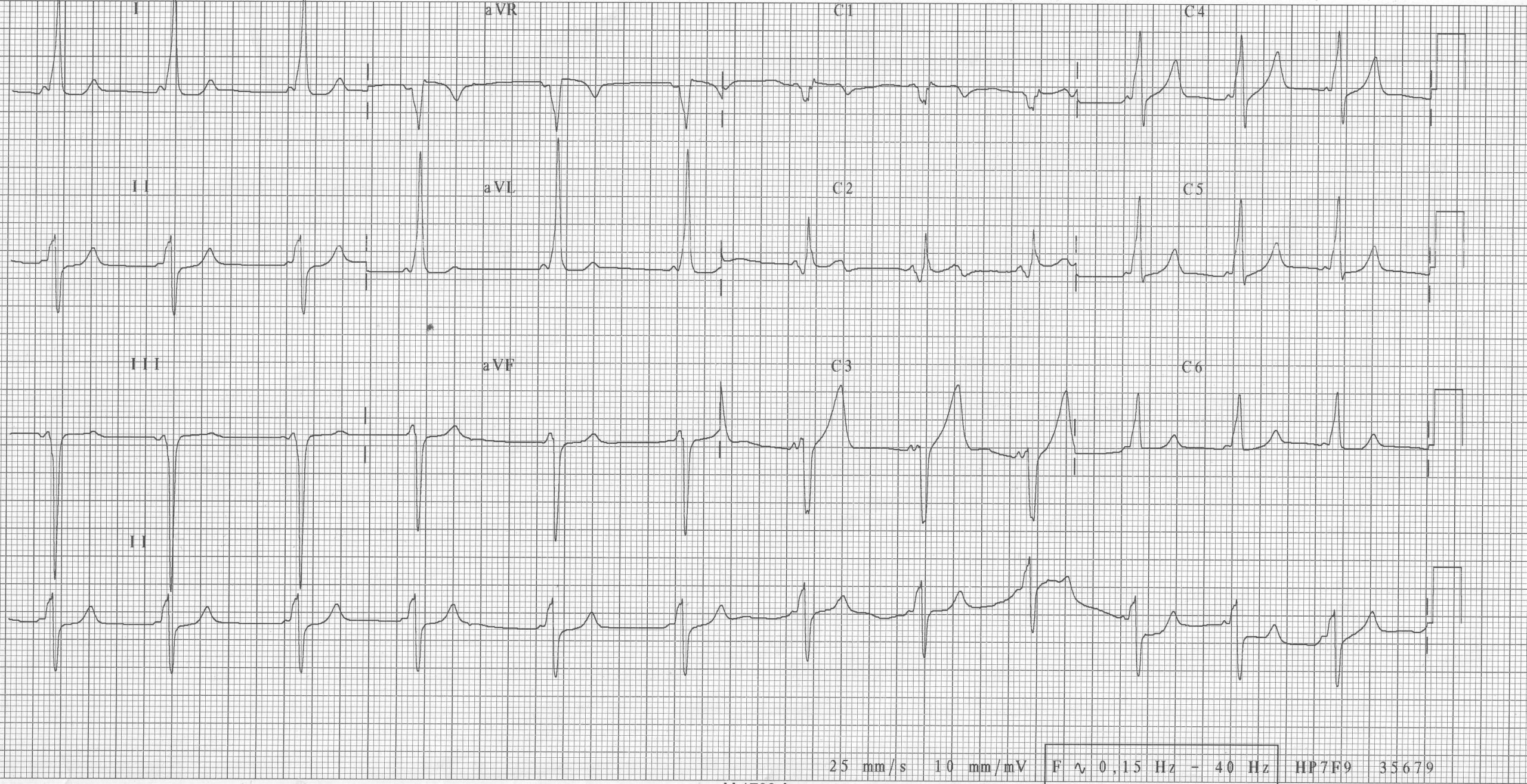
Ebstein’s disease is classically characterized by an abnormal development of the tricuspid valve, the septal leaf being displaced into the right ventricle. A surgical correction is often necessary in the presence of significant valvular regurgitation, or an associated cyanosis (by inter-atrial shunt) and/or other associated anatomical abnormalities. In adolescence and adulthood, disease evolution is marked by the common occurrence of arrhythmia episodes.
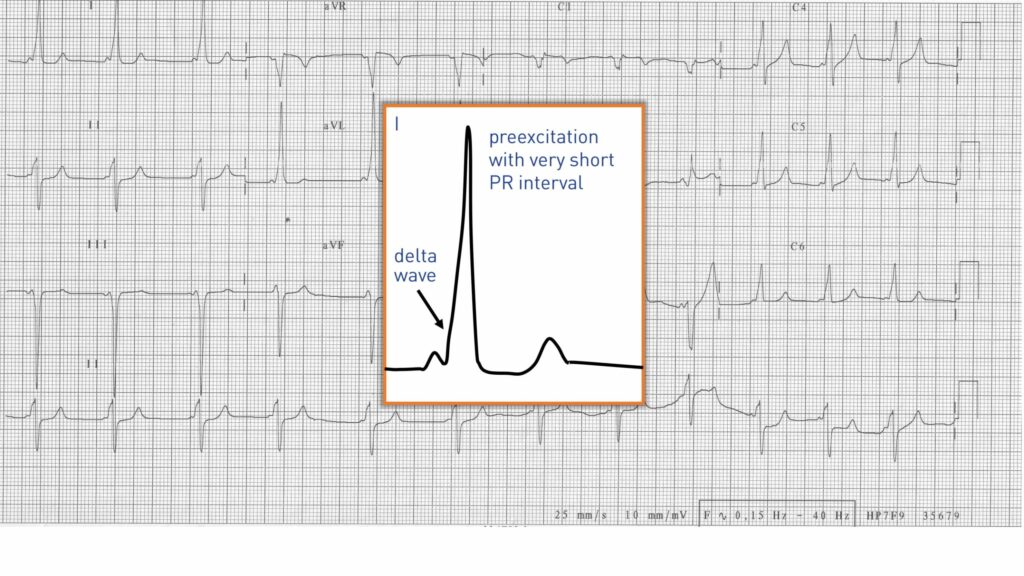
Ebstein’s disease is a congenital heart disease where presence of an accessory pathway is the most common observation since estimated between 10 and 40% (versus less than 0.1% in the general population). The vast majority of these accessory pathways are located on the right and primarily at the level of the posteroseptal or posterolateral aspect of the tricuspid annulus. The diagnosis is sometimes difficult to make on the electrocardiogram. Indeed, a major intra-atrial conduction delay is frequent resulting in a prolonged period between sinus node and atrial portion of the accessory pathway. These accessory pathways are often long and have slow conduction velocities. This results in a longer PR than for a traditional accessory pathway and a minimally pre-excited QRS complex. This is not the case in this patient where the PR interval is very short and the pre-excitation easily visualized. In addition, a considerable number of patients exhibit several accessory pathways that render localization on the electrocardiogram difficult.
It is possible, as in this patient, to highlight certain electrocardiographic characteristics. Pre-excitation suppresses the typical pattern of right bundle branch block observed in this type of heart disease, with in general a left axis deviation of the QRS in the frontal plane and QS or rS morphology in V1. The absence of a right bundle branch block on the surface electrocardiogram should therefore suggest the presence of an accessory pathway. Similarly, the reappearance of a right bundle branch block pattern after ablation suggests the effectiveness of the procedure.
English English English French English English English French