


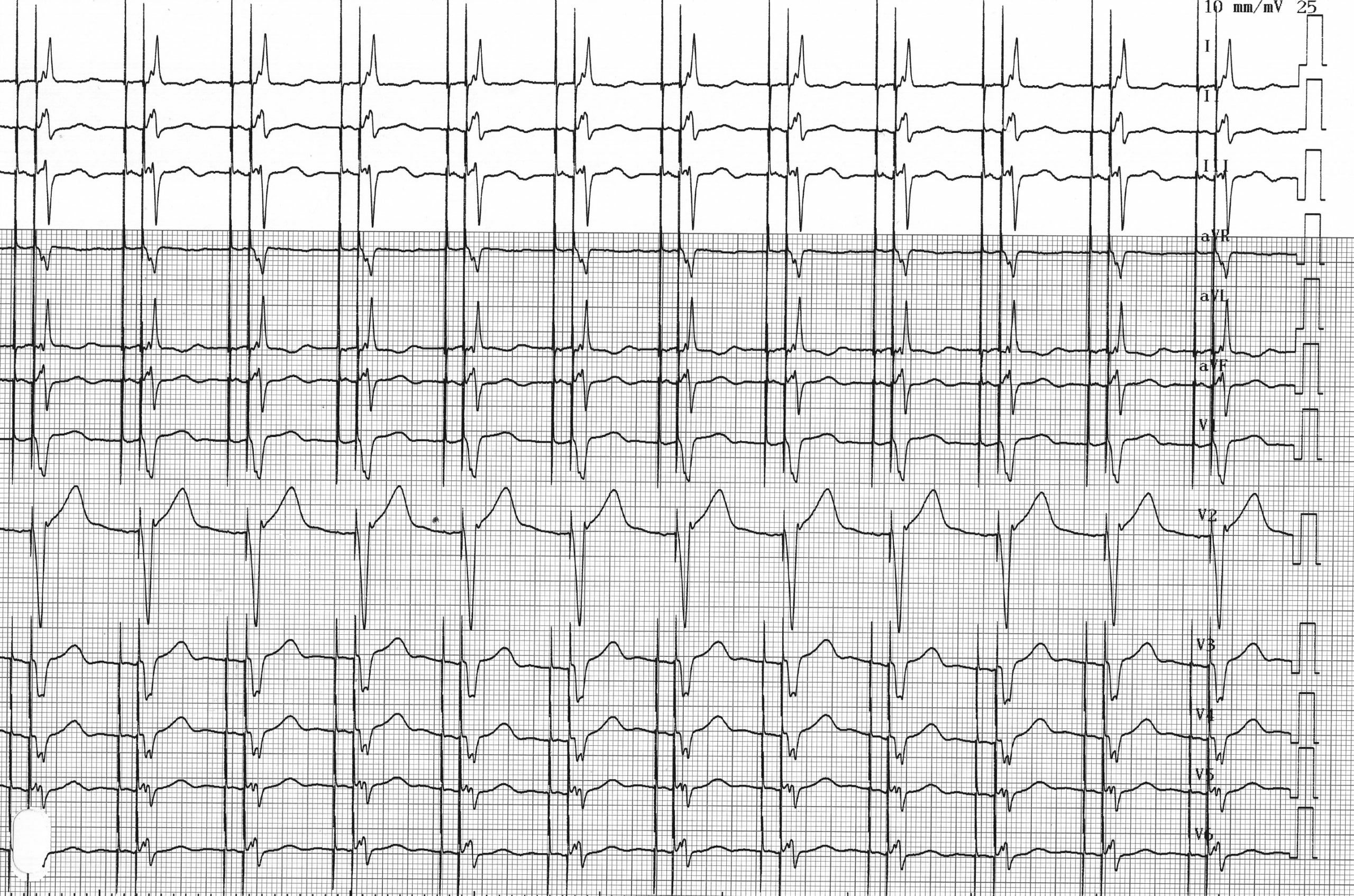
DDD mode is the standard programming mode for dual-chamber or resynchronization pacemakers. It ensures atrioventricular synchronization at rest and during exercise based on detected or stimulated atrial activity. In fact, the basic principle of DDD mode is to synchronize ventricular pacing with detected (triggered) or stimulated atrial activity. Spontaneous atrial or ventricular activity detected outside the refractory period inhibits atrial or ventricular pacing, respectively (inhibited mode). This mode therefore maintains atrioventricular synchrony across a wide range of sinus rates, from low to high (up to the maximum rate limit). Any atrial detection outside the refractory period between the base rate and the maximum synchronous rate, or atrial pacing, results in an AV delay with ventricular pacing in the absence of spontaneous ventricular detection. The AV delays following detected atrial events and stimulated atrial events are programmable separately; it is possible to program the AV delays to shorten as rates increase (adaptive AV delay) or to adjust them based on spontaneous conduction times.
In DDDR mode, the pacemaker follows the faster of the two rates: the intrinsic atrial rate or the rate indicated by the sensor. The maximum synchronous rate and the maximum slave rate can be programmed separately.
The DDD mode programming therefore appears to be adequate for this patient. However, follow-up monitoring would likely reveal a ventricular pacing percentage close to 100%. This would therefore correspond to functioning that is considered normal and appropriate. The 0D0 analysis, however, revealed that this patient has normal AV conduction. One of the priorities in pacemaker programming is to avoid any unnecessary right ventricular pacing.
This helps reduce battery consumption and extend the device’s lifespan and, most importantly, avoid right ventricular pacing, which is associated in the short, medium, and long term with adverse effects on hemodynamics, ventricular remodeling, and the onset of atrial arrhythmias. In fact, right ventricular pacing induces an asynchronous sequence of interventricular and intraventricular activation and relaxation.
A high percentage of ventricular pacing in a patient with preserved AV conduction should raise the clinician’s suspicion during the medical history and prompt a discussion of the use of specific algorithms that promote spontaneous conduction.
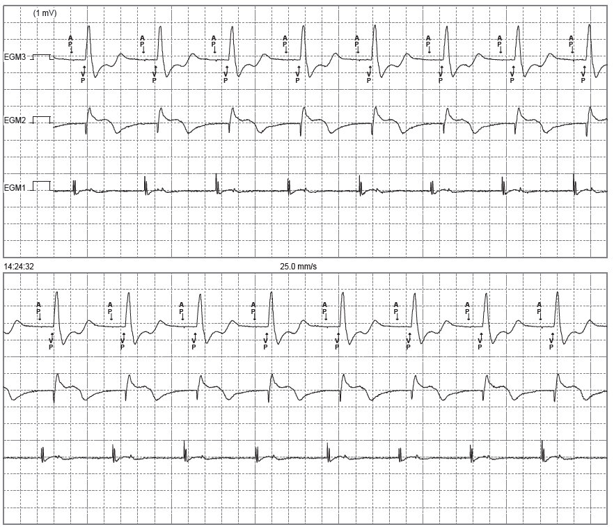
Trace description
AP-VP cycle repetition (atrial and ventricular pacing) at 70 bpm;
English English English French English English English French