


Microport - Oversensing
Content
Ventricular sensitivity control
1 - Principles
The purpose of the Automatic Ventricular Sensitivity Control function is to improve sensing of ventricular fibrillation (VF): a high sensitivity of the device must be applied during VF since most VF signals are of low amplitude (often amplitudes less than 3 mV), and also to avoid sensing of the T wave both in sinus rhythm and during arrhythmias.
One of the challenges of improving VF detection is that the signal amplitude is much lower during VF than in sinus rhythm and the difference in amplitude from one beat to the next can vary.
Another challenge is to avoid sensing of T waves, the amplitude of the filtered T waves is generally less than that of the R waves (the amplitude of the T wave is of the order of 25% of the R amplitude). Pre-clinical studies have shown that the maximum amplitude of these T waves is less than 25% of R waves after filtering.
2 - Filtering
The system used in pacemakers and cardiac defibrillators for the selection or non-selection of a signal is based on filtering techniques. All devices use amplifiers designed to detect events whose frequency varies between an upper limit and a lower limit: the bandwidth. Any event whose frequency is outside these limits will not be taken into account by the device. For MicroPort defibrillators, the ICD bandwidth is 25 to 80 Hz (at -3 dB), and is centered at 55 Hz. Filtering thus allows discriminating T waves, R waves or PACs. On the other hand, given that the filter limits are not completely vertical, it is possible that the circuit detects events outside its bandwidth, if their amplitudes are sufficiently significant. A classic but rare example of this type of oversensing is the sensing and recognition by the defibrillator of the T wave as being an R wave (recycling on the T wave).
3 - Ventricular sensing
For the Automatic Sensitivity Control function, only R waves are filtered and adjusted before being analysed for detection. After filtering, the T waves are attenuated and the signal obtained is rendered positive, in order to detect positive and negative slopes.
At first, a non-programmable absolute refractory period of 95 ms is applied after the sensing of the R-wave.
During the first 64 ms of this period, the implant still measures the amplitude of the R wave. The maximum amplitude of the R waves that the implant can measure is capped at 6 mV for this function.
For the ongoing cycle, the sensitivity will change as a function of the amplitude of the R wave measured in the first 64 ms. It can never go below the programmed sensitivity.
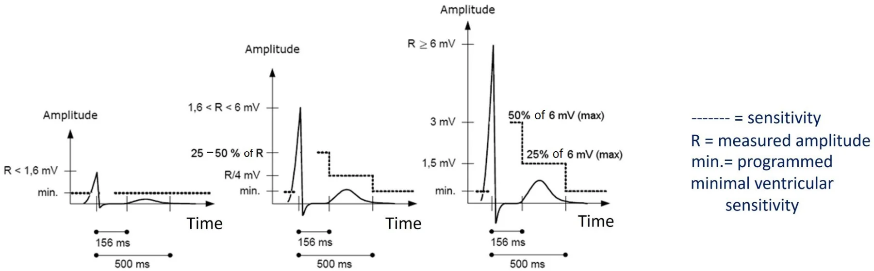
During the 95-156 ms interval following the sensing of the R wave:
- If the amplitude of the sensed R wave was greater than 6 mV, a sensitivity of 3 mV is applied (3 mV = 50% of 6 mV),
- If the amplitude of the sensed R wave was less than 1.6 mV, a sensitivity of 0.4 mV (the programmed sensitivity) is applied (0.4 mV = 25% of 1.6 mV),
- Between 1.6 and 6 mV, the applied sensitivity is between 25 and 50% of the measured R-wave. The sensitivity is calculated linearly in this interval: [1.6 mV; 6 mV] -> [25%; 50%].
These settings allow avoiding the multiple sensing of wide R waves and to sense the R waves in VF.
In the 156-500 ms interval following the sensing of the R wave, the sensitivity is set at 25% of the amplitude of the measured R wave (never below the programmed sensitivity). This setting was implemented to reject the T waves.
The following table illustrates how the device sets the sensitivity value for the 2 plateaux proportionally to the amplitude of the measured R wave. The greater the amplitude of the R wave, the lower the sensitivity of the device (i.e. high sensitivity value).
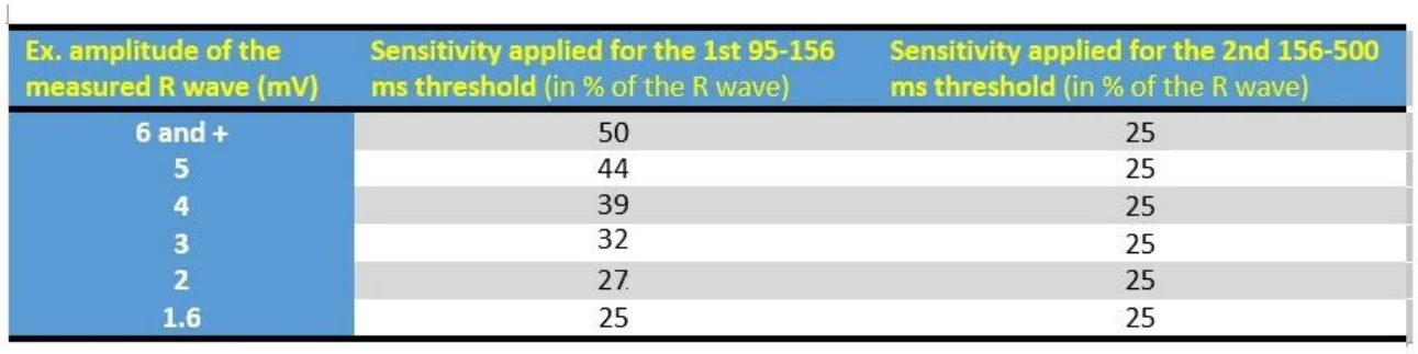
Thus, during much of the cycle (after 156 ms), ventricular sensitivity is comprised between 1.5 mV and the programmed sensitivity value in the event of a complex measured at an amplitude greater than 4 mV.
After 500 ms following the sensing of the R wave, and until the end of the cycle, the sensitivity returns to the programmed value or to 1mV (not configurable) according to certain conditions.
If all of the following conditions are fulfilled:
- the programmed ventricular sensitivity value is less than 1 mV, and
- the device does not detect a short cycle (VF/Fast VT zone) over the last six cycles, and
- the measured amplitude is greater than or equal to 4 mV
then, the V detection threshold is set at 1 mV after the second threshold; i.e. after 500 ms.
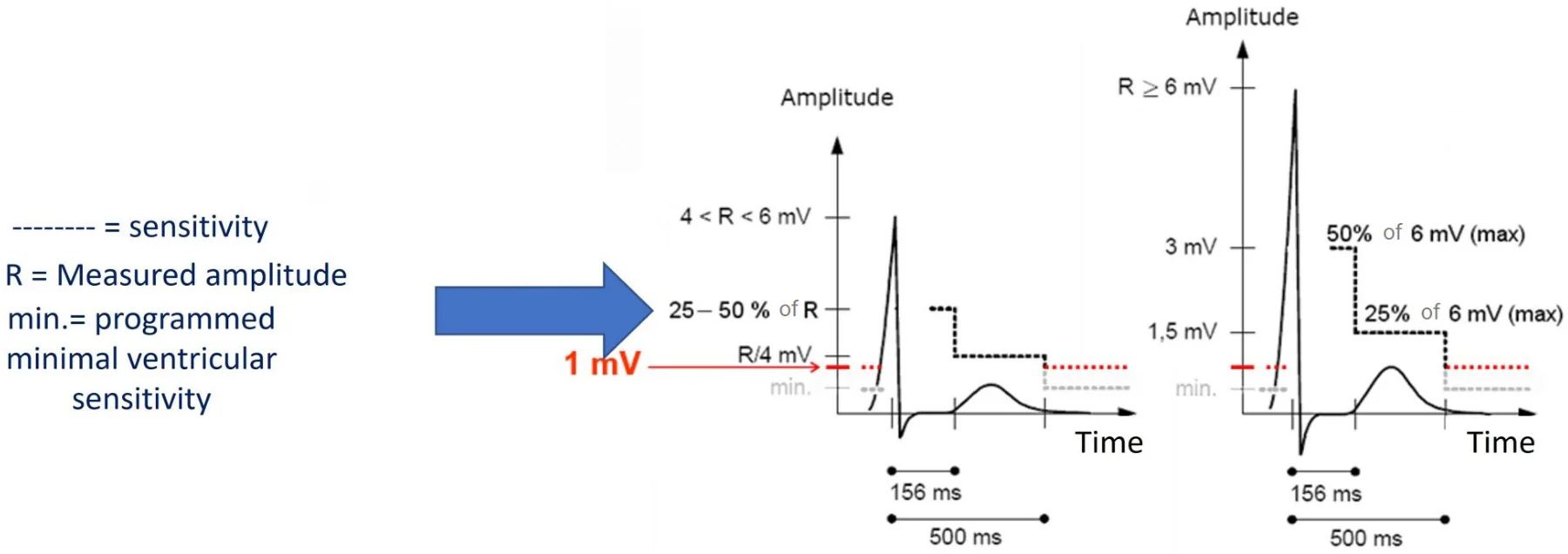
In sinus rhythm and in the VT zone, the lowest detection limit will be 1 mV and will cover any background noise (myopotential). During this period, the V sensitivity will mostly be 1 mV.
In case of a short coupling interval or weak detection, the lowest detection threshold will decrease to the programmed sensitivity (0.4 mV nominal).
If one of the following conditions is fulfilled:
- the device detects at least one short cycle (VF/Fast VT zone) over the last six cycles, or
- the measured R-wave amplitude is less than 4 mV (not applicable for the 3rd diagram)
then, the V detection threshold is set to the programmed sensitivity after the second plateau; i.e. after 500 ms.
4 - Ventricular pacing
After ventricular pacing, there is an “automatic” management of the sensitivity. At first, an absolute non-programmable 220 msec refractory period is applied after the ventricular pacing. An additional sensitivity margin is programmable (nominal 0.8 mV) for the period between 220 ms and 500 ms post-ventricular pacing, this is the “V margin Post-V pacing”. This is a value added to the programmed ventricular sensitivity for this period. The goal is to “cover” broader T-waves in the case of ventricular pacing in general. After this 500 ms period, ventricular sensitivity automatically returns to 1 mV or the programmed sensitivity if the following conditions are fulfilled:
- the V programmed sensitivity is programmed > 1 mV, and
- if one or more short cycle (cycle in Fast VT/VF zone) was detected over the last six cycles
then the detection threshold V is set at 1 mV after 500 ms.
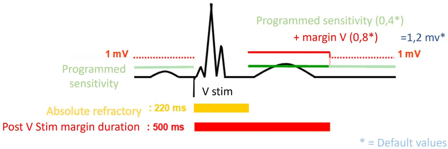
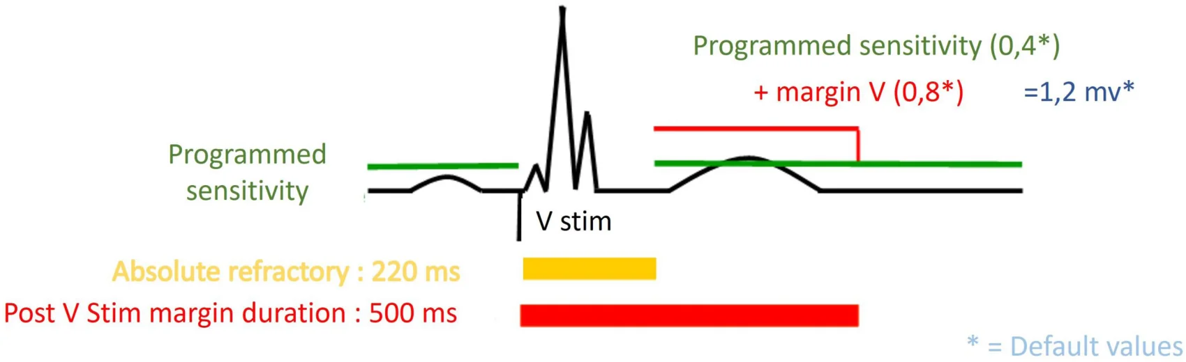
If there is at least one short cycle (fast VT/VF zone) in the last six cycles, then the V detection threshold is set to the programmed sensitivity after 500 ms.
5 - Programming
The Automatic Sensitivity Control is fully automatic. In case of difficulty linked to ventricular detection, the only parameter that can be adjusted is ventricular sensitivity.
There is only one possible setting in the case of ventricular pacing. This is the “V margin post-V pacing” parameter. See the chapter on automatic management of post-ventricular pacing sensitivity. You will need to open the “Expert parameters” and choose “A and V Detection”. The “V Margin post-V pacing” parameter is then accessible. By default, knowing that its value is 0.8 mV and the ventricular sensitivity is 0.4 mV, the applied post-ventricular pacing ventricular sensitivity for the period from 220 ms to 500 ms is 1.2 mV.
Atrial sensitivity control
The control of atrial sensitivity enables avoiding R-wave crosstalk (or far-field sensing), both in sinus rhythm and during tachycardias. It is coupled with an automatic prolongation of the refractory periods after ventricular events. The 2 strategies are often combined.
1 - Implemented strategies for managing crosstalk of ventricular activity in the atrial channel
Since the atrial channel has a high nominal sensitivity (0.4 mV programmed sensitivity) and by definition short refractory periods, the sensing quality on this channel has been put to the test particularly in situations of crosstalk or far-field sensing (R or V sensing in the A channel). Different solutions have been studied and applied to improve sensing on the atrial channel.
2 - Programming
Several settings are available to improve the quality of atrial sensing. Two parameters affect the value of the applied A sensitivity: “A Sensitivity” and “A margin post R or V”. Two parameters affect the duration of the absolute atrial refractory period: the “Post V Atrial Refractory Period” and the “Post R Atrial Refractory Period”.
3 - Situation where the ventricular event is preceded by atrial sensing
- In the case of ventricular detection preceded by atrial sensing, after the absolute refractory period (45 ms), the value of the applied sensitivity is the programmed sensitivity (0.4 mV) incremented by the “A margin Post R or V” (0.4 mV) for 30 ms* + 97 ms
- In the case of ventricular pacing preceded by atrial sensing, after the absolute refractory period (80 ms), the value of the applied sensitivity is the programmed sensitivity (0.4 mV) incremented by the “A margin Post R or V” (0.4 mV) for 30 ms* + 62 ms.
* Re-triggerable relative refractory period.
4 - Situation of PVC
- In the case of a PVC, after the absolute refractory period (45 ms), the value of the applied sensitivity is the programmed sensitivity (0.4 mV) incremented by the “A Margin post R or V” (0.4 mV) for 30 ms* + 97 ms.
5 - Situation where the ventricular event is preceded by atrial pacing
- In the case of ventricular detection preceded by atrial pacing, the absolute refractory period (45 ms) is prolonged by 65 ms and by the re-triggerable relative refractory period (30 ms).
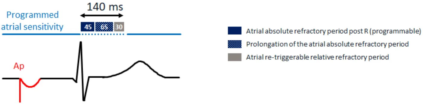
Note: In AAI-SafeR mode, in the case of ventricular detection preceded by atrial pacing, the absolute atrial refractory periods are not prolonged and the atrial sensitivity margin is not added. Only the absolute refractory period (45 ms) prolonged by the re-triggerable relative refractory period (30 ms) is applied.
- In the case of ventricular pacing, preceded by atrial pacing, the absolute refractory period (80 ms) is prolonged by 65 ms and by the re-triggerable relative refractory period (30 ms). The “A sensitivity control” is carried out over a total of 175 ms (80+65+30) after the sensing of the R-wave.
Oversensing situations
An oversensing of events detected by the defibrillation ventricular lead can occur and is added to the normal sensing of the R-wave. This results in false diagnoses of ventricular arrhythmia with the possible triggering of inappropriate and dangerous therapies. (At the atrial level, oversensing can also lead to either a false diagnosis of atrial arrhythmia and thus an inappropriate mode switch, or an additional degree of artificial PR association which, concomitant with a stable and dissociated VT, leads to a diagnosis of SVT).
There are two types of oversensing: those that are intrinsic, via the sensing of cardiac events of atrial origin (P waves, atrial tachycardia waves) or of ventricular origin (double sensing of R waves, T waves), and those that are extrinsic, via the sensing of non-cardiac bodily potentials (pectoral or diaphragmatic myopotentials), or lead fracture potentials, or extracorporeal potentials (external or fortuitous interference (50 Hz line, for example), or by the use of medical equipment (magnetic or electric or electromagnetic field of a diagnostic or therapy apparatus (electrotherapy, diathermy knife, for example)).
In all instances, the number of detected events per cardiac cycle is in excess and will result in two consequences: i) a consequence on cardiac pacing with an inhibition in pacing which, in a device-dependent patient, leads to a cardiac pause and its ensuing clinical course as a function of the duration of the pause; ii) a consequence on defibrillation, with false diagnoses of ventricular arrhythmia leading to unexpected shocks, which in turn can trigger true cardiac arrhythmias. Shocks are often iterative as long as the cause of this oversensing has not been removed. The use of a magnet should thus be reminded and whose action will be to suspend any detection of arrhythmia and all therapies as long as it applied next to the defibrillator case.
The suspicion of an oversensing-type activity, whether intrinsic or extrinsic, is evoked by the pattern exhibited by the tachogram of the recorded episode.
Intrinsic oversensing
The sensing of P or T waves, or the double counting of R waves will cause oversensing for fixed intervals relative to the QRS detection signal, corresponding to the P or T wave onset times, or an intra-QRS interval. This results in a characteristic « railroad track » pattern which quite easily evokes this type of artifact whose type will be detailed by the electrograms. Clinically, in addition to the consequences of untimely triggered therapies in the absence of any arrhythmia, such oversensing at the ventricular level incurs a recycling of the pacemaker, and in a bradycardia patient, the pacing rate will be slow on the surface ECG, i.e. slower than what should normally be observed.
For more information on Oversensing in MicroPort ICDs, please see: 7. Oversensing – MicroPort Academy.