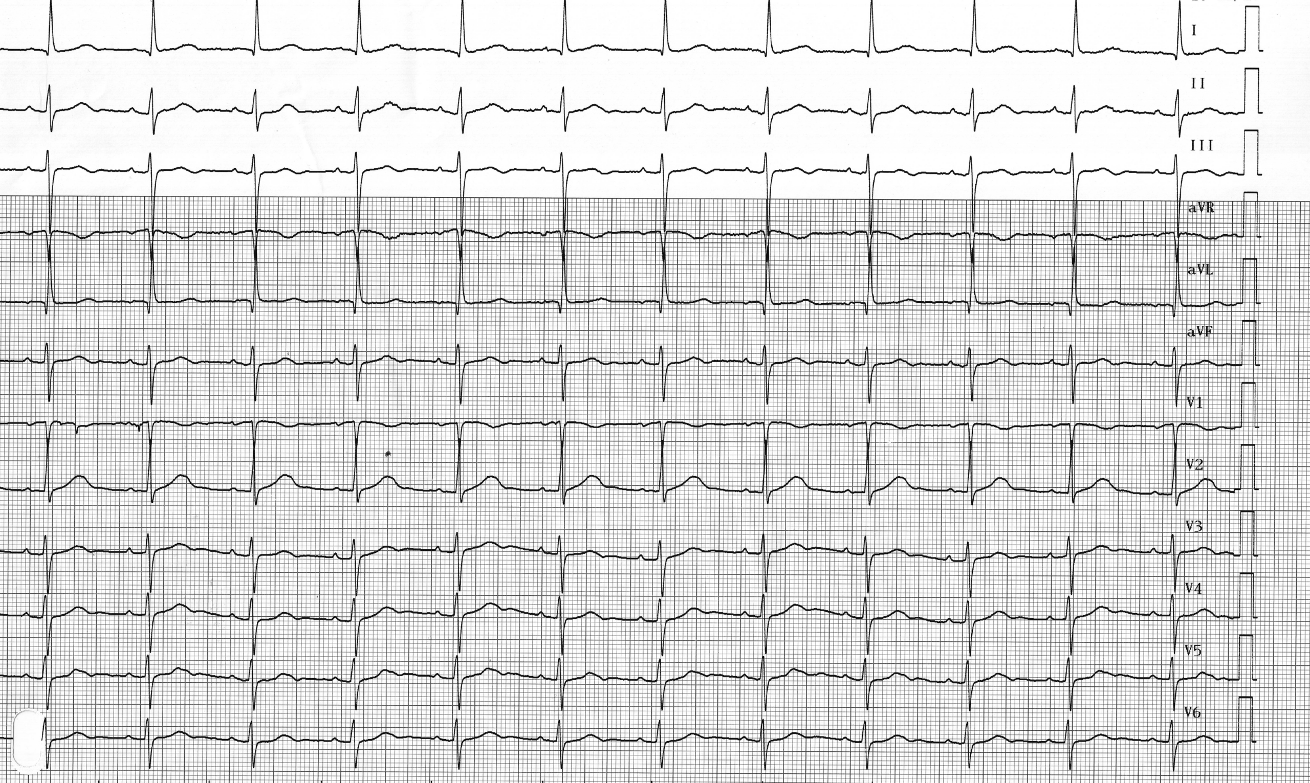
A 69-year-old man who received a Medtronic dual-chamber pacemaker due to recurrent syncope caused by sinus node dysfunction; both leads are well-positioned with normal pacing impedances, correct detection, and satisfactory pacing thresholds; Three days after implantation, the pacemaker was checked; various pacing modes were programmed, and tracings were recorded; for this first tracing, the device was programmed to 0D0 mode
Regarding this ECG, which of the following is/are the correct answer(s)?
ECG description
Spontaneous atrial contractions and sinus rhythm (positive P waves in DI, DII, V5, V6; negative in aVR); normal, fixed PR interval (< 200 ms); spontaneous ventricular contractions with signs of left anterior hemiblock (left axis, q1S3 pattern);
Explanation
Depending on the number of leads and the model of the implanted device, different stimulation modes can be programmed. The treating physician must understand the operation, advantages, and disadvantages specific to each stimulation mode in order to optimize programming based on the individual characteristics of each implanted patient. A collaborative effort between the North American Society for Pacing and Electrophysiology (NASPE) and the British Pacing and Electrophysiology Group (BPEG) led to the development of an international classification code (NBG) for the various pacing modes. The various stimulation modes are identified by a 4-letter code that describes their basic operation. The first letter defines the stimulation site(s): ventricle (V), atrium (A), both (D), single chamber (S), or none (0). The second letter defines the detection site(s): the same letters as above. The third letter indicates the operating mode: inhibited (I), triggered (T), both (D), or none of the above (0). The fourth letter indicates rate response (R) or its absence (0).
In this first trace, the pacemaker was programmed to 0D0 mode. The distinctive feature of this “stimulation” mode is that detection in both chambers is active, but no stimulation (atrial or ventricular) is possible. It should therefore obviously not be used in dependent patients. This pacing mode thus allows for analysis of the patient’s spontaneous rhythm with simultaneous visualization of the electrocardiogram and event markers. It is a sensitivity test mode. The 0D0 mode can be temporarily programmed in non-dependent patients with an MRI-compatible pacemaker who are scheduled to undergo an MRI. The 0D0 mode of these MRI-compatible pacemakers prevents the potential reversion from a traditional 0D0 mode to a VVI mode. It is, of course, essential to check and reprogram the pacemaker after the exam (according to the manufacturer’s instructions).
In the following waveforms, various markers are used:
AS (atrial sensing): atrial sensing; VS (ventricular sensing): ventricular sensing; AP (atrial pacing): atrial pacing; VP (ventricular pacing): ventricular pacing;
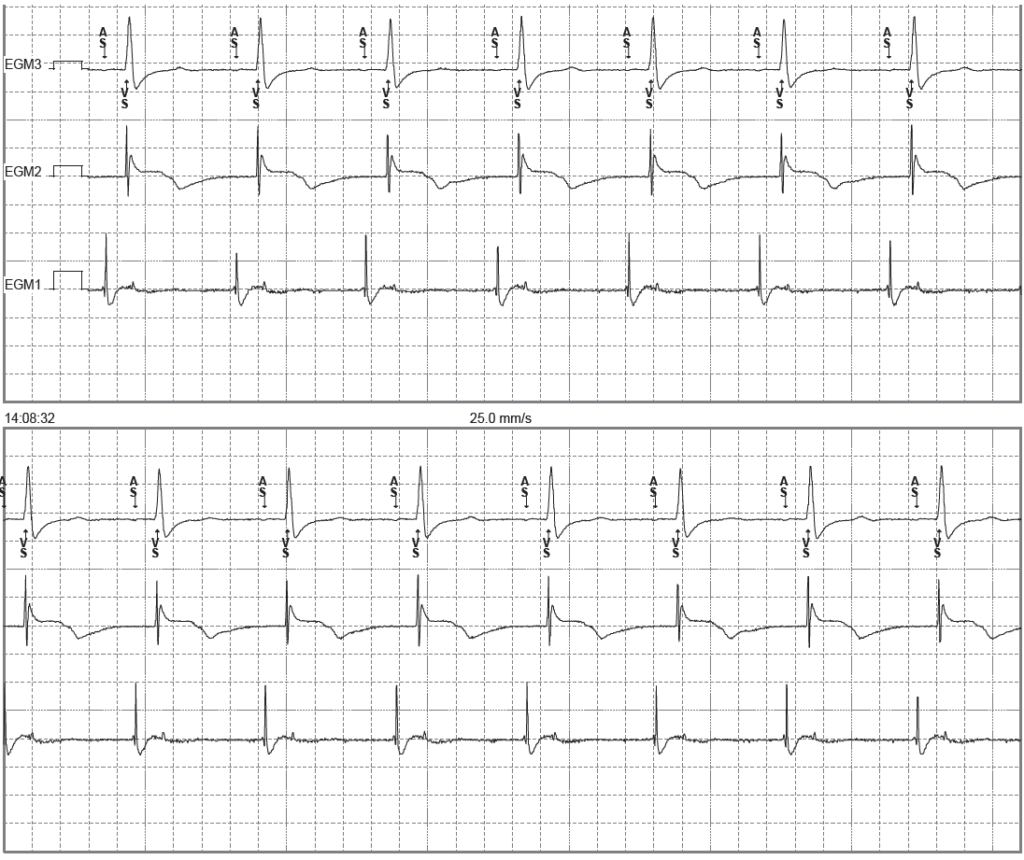
Trace description
This refers to the recording (simultaneously with the previous trace) of intracavitary electrograms from the programmer; EGM 3 corresponds to an electrocardiographic lead, EGM 2 to the ventricular EGM, and EGM 1 to the atrial EGM; spontaneous atrial and ventricular activity (AS-VS);
To remember
On a dual-chamber pacemaker, various modes can be programmed, each with its own specific characteristics; D00 mode: no sensing capability, asynchronous pacing; mode 0D0: no pacing; mode DDT: triggered mode, where a detection triggers a pacing pulse; mode VDD: no atrial pacing; mode DDI: no ventricular pacing following an atrial detection; mode DDD: ventricular synchronization with atrial detection and ventricular pacing; manufacturer-specific modes to prevent unnecessary ventricular pacing;