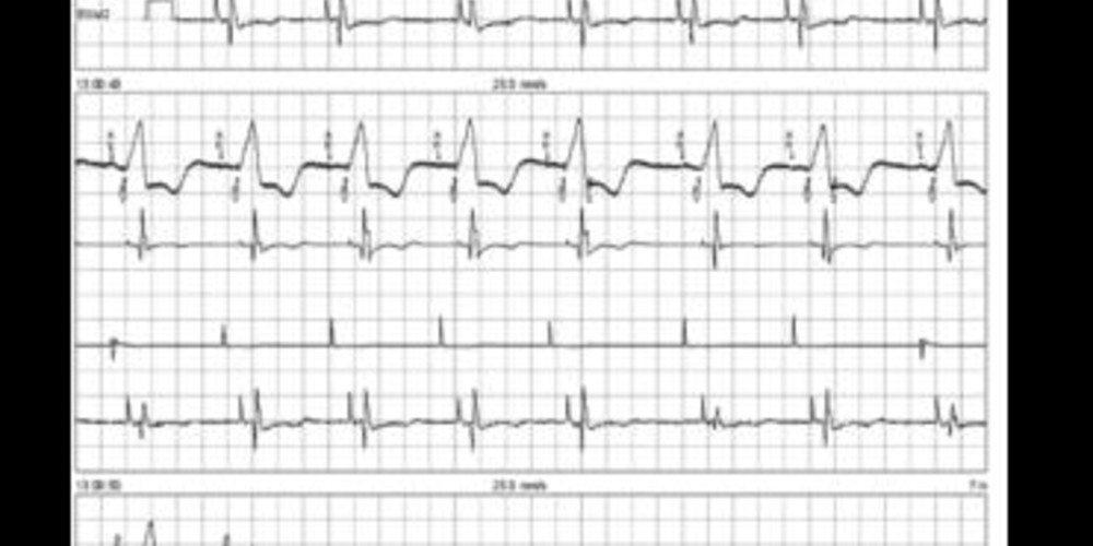
Loss of RV capture
Tracing
Manufacturer Medtronic
Device CRT
Field Left ventricular pacing
N° 7
Patient
70-year-old man implanted with a triple chamber defibrillator Viva XT CRT-D for severe ischemic cardiomyopathy and left bundle branch block; good response to resynchronization therapy; two endocarditis episodes necessitated the complete removal of the system (first implanted on the left side, then on the right side); complete surgical re-implantation of a defibrillator with 2 coils, a bipolar atrial and RV lead and a bipolar LV lead; routine follow-up.

Graph and trace
- atrial and biventricular pacing (AP-BV);
- double counting of the paced QRS (BV-FS);
RV pacing threshold test performed in bipolar configuration (DDD mode, 90 bpm) - first pattern in RV pacing (probable double anodal and cathodal capture of the right cavity);
- change in the paced QRS pattern (pure cathodal capture);
- loss of RV capture (threshold 5.5 volts/0.7 ms);
- biventricular pacing with an amplitude of 5 volts/0.7 ms;
- programming change with an increase of the amplitude to 8 volts (superior to the RV threshold);
- clear modification of the QRS pattern with a narrowing of the QRS and biventricular capture.
Other articles that may be of interest to you


Traditionally, the loss of right ventricular capture by dislodgement or by threshold elevation is a rarer occurrence than the loss of left ventricular capture, the right lead being screwed into the endocardium and the left lead deposited at the epicardium. In this patient, the right ventricular lead was implanted surgically. The two electrodes are sewn onto the epicardium and are relatively distant, which explains the increase in threshold (epicardial thresholds often higher) but also the double QRS pattern. The thresholds are readily elevated after surgical implantation before a clear trend toward improvement during the first 3 months, a phenomenon favored by steroid elution (4968 leads).
The loss of right ventricular capture is accompanied by a lone left ventricular capture without adaptation of refractory periods or post-ventricular pace ventricular blanking. Left ventricular pacing is often associated with a widened QRS and an increased risk of double counting of the paced QRS sensed by the right ventricular lead. In this patient, a left ventricular pacing configuration was favored with programming of a post-ventricular pacing ventricular blanking of 220 ms. This long blanking period may potentially delay the sensing of the first arrhythmic cycle during an episode of VF but does not compromise its sensing in a prolonged manner, the device switching to the ventricular post-sense blanking following the sensing of a spontaneous ventricle.