Dual chamber and V< A branch discrimination
Patient
Go to the casebook version optimized for the mobile experience
Simulator-generated 2:1 conducted atrial flutter trace on a dual-chamber Gallant defibrillator
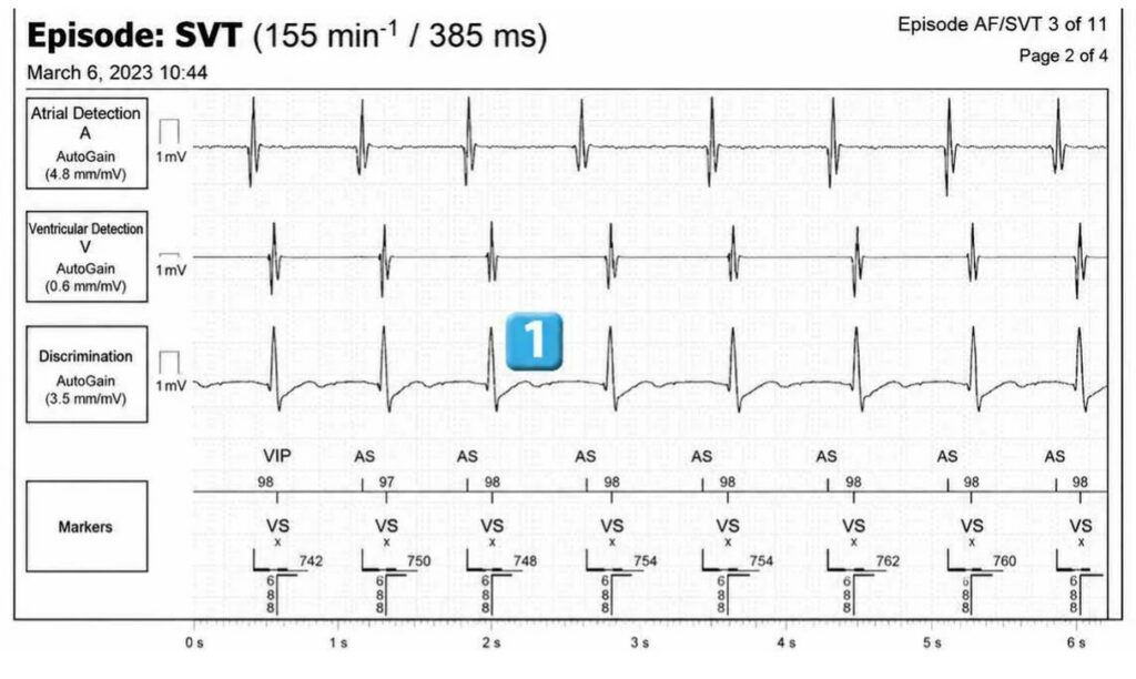
Trace
Episode 1
- Atrial detection and ventricular sensing
- onset of rapid, polymorphic atrial arrhythmia (AF)
- cycles classified as T1; most ventricular cycles have a correlation percentage above the programmed threshold
- after 3 cycles classified as T1, switch to episode mode (VVI), atrial markers are no longer displayed on the trace
- VT1 counter completed; SVT diagnosis (in branch V< A)
- after 6 cycles classified as T1, SVT redetection
- spontaneous reduction on a ventricular complex
- diagnosis of end of episode
Episode 2
- Atrial detection and ventricular sensing
- onset of monomorphic, regular atrial arrhythmia; 2:1 atrioventricular conduction
- cycles classified as T1; most ventricular cycles have a correlation percentage greater than the programmed threshold
- after 3 cycles classified as T1, switch to episode mode (VVI), atrial markers are no longer displayed on the trace
- VT1 counter completed; SVT diagnosis (in the V< A branch); tachycardia is stable at the ventricular level with stable A/V coupling
- after 6 cycles classified as T1, SVT redetection
- spontaneous reduction on a ventricular complex
- diagnosis of end of episode
Comments
SVT in the V<A branch The two discriminating factors (morphology and interval stability) favor a SVT
SVT in the V<A branch Both discriminants (morphology and interval stability) are consistent with SVT
Points to remember
- In the presence of tachycardia in the V<A branch, various diagnoses are possible (bitachycardia, conducted AF, conducted atrial flutter/tachycardia).
- In the V<A branch, various discriminating factors are used: morphology, stability/ AV association
- In the case of bitachycardia (VT+AF/flutter), the morphology is different in tachycardia
- compared to sinus rhythm; the ventricular rhythm is stable with no association with the atrial rhythm.
- In conducted AF, the morphology is identical in tachycardia compared to sinus rhythm; the ventricular rhythm is unstable.
- During conducted atrial flutter/tachycardia, the morphology is identical in tachycardia compared to sinus rhythm; ventricular rhythm is stable with AV association
- The “All” setting is more specific than the “One of” setting.
- AV coupling works on the same principle as frequency stability. The AV coupling delta measures the difference between the second longest AV interval and the second shortest AV interval among the 12 cycles (programmable) preceding detection. If the measured difference is less than the programmed delta, then ventricular rhythm is associated, and the parameter favors SVT (flutter or atrial tachycardia conducted in n/1). Otherwise, the parameter favors VT is associated, and the parameter favors SVT (flutter or atrial tachycardia conducted in n/1) otherwise, the parameter favors VT. This criterion only applies if the rhythm is stable, and the setting is common: stability programmed to On + AVA
Read more about related topics