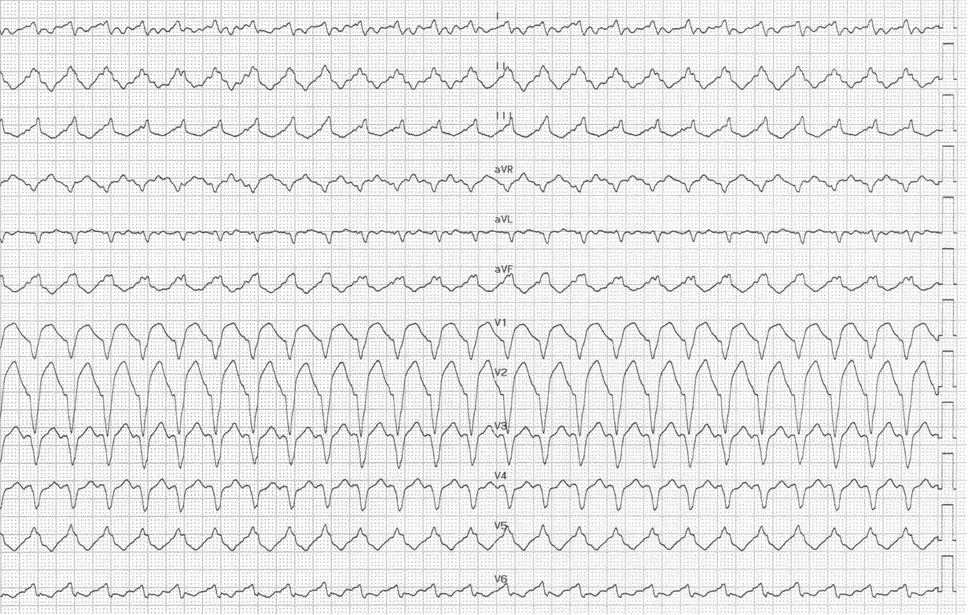
Tracé 8.5 : Tachycardie ventriculaire avec bloc de branche
Informations destinées aux patients
Patient âgé de 74 ans, atteint de fibrillation auriculaire permanente, ayant subi une ablation du nœud auriculo-ventriculaire et la pose d'un stimulateur cardiaque à trois chambres (sondes de stimulation ventriculaire droite et gauche) ; hospitalisation pour un épisode syncopal ;
ECG
Question
Compte tenu des antécédents cliniques du patient et de son tracé ECG, quel(s) est (sont) le(s) diagnostic(s) possible(s) pour cet ECG ?
Commentaires
Description de l'ECG
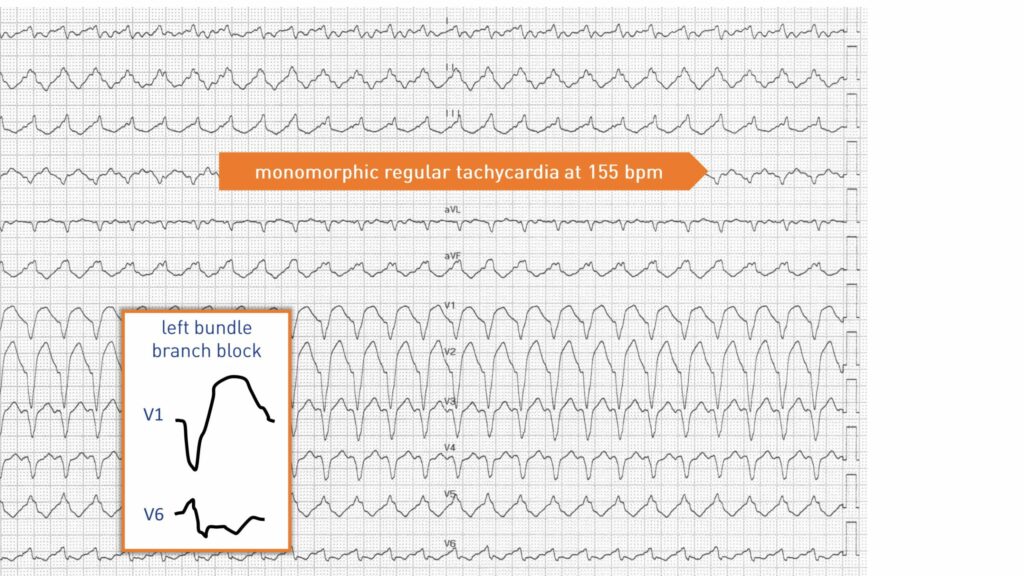
Ce tracé montre une tachycardie monomorphe et régulière à 155 bpm, avec un tracé caractéristique d’un bloc de branche gauche ; l’activité auriculaire est difficile à identifier ; les antécédents cliniques indiquent que ce patient a subi une ablation du faisceau de His et présente donc un bloc auriculo-ventriculaire antérograde complet ; le seul diagnostic possible en présence d'une tachycardie avec complexes QRS spontanés (absence de stimulation ventriculaire) est donc une tachycardie ventriculaire ; les antécédents cliniques peuvent parfois influencer l'interprétation d'un tracé (des antécédents d'infarctus suggèrent une tachycardie ventriculaire mais ne garantissent pas son diagnostic) mais peuvent parfois permettre un diagnostic certain (comme dans ce cas où il existe un bloc auriculo-ventriculaire complet) ; tachycardie de réentrée probable de branche à branche ;
Explication
La réentrée branche-à-branche est une tachycardie ventriculaire résultant d’une macro-réentrée entre les deux branches. Ce type de tachycardie représente un pourcentage significatif (de l’ordre de 20 à 40%) des tachycardies ventriculaires observées chez les patients atteints de cardiomyopathie idiopathique non ischémique. Un certain nombre de cas ont également été décrits dans le cadre de cardiomyopathies valvulaires ou ischémiques, chez des patients atteints de myotonie et chez certains patients ne présentant pas de maladie cardiaque. La survenue de quasi-syncopes, de syncopes ou de morts subites est fréquente, car la tachycardie est souvent rapide et survient en présence d’altérations myocardiques. La branche droite constitue la voie antérograde dans la majorité des cas, l’un des faisceaux (antérieur ou postérieur) de la branche gauche constituant la voie rétrograde, ce qui explique le tracé de bloc de branche gauche. Le circuit inverse (schéma de bloc de branche droit) est plus rare. Le déclenchement de la réentrée se produit lorsqu’un complexe auriculaire ou ventriculaire prématuré est bloqué dans l’une des branches, conduit dans l’autre en direction antérograde, puis pénètre dans la branche initialement bloquée de manière rétrograde, induisant une tachycardie de réentrée soutenue. L'électrocardiogramme en rythme sinusal révèle fréquemment un bloc auriculo-ventriculaire du premier degré, un trouble de conduction distal non spécifique, rarement accompagné d'un bloc de branche gauche. La condition préalable à l’apparition d’une réentrée branche-à-branche est la présence d’un retard de conduction dans le système de His-Purkinje, se traduisant par un intervalle HV prolongé (80 ms en moyenne). Chez ce patient, l’ablation du nœud auriculo-ventriculaire ne permet pas d’analyser la séquence d’activation ventriculaire spontanée. Les traitements antiarythmiques sont rarement efficaces dans ce contexte. L’ablation de la branche droite résout souvent le problème, même si un défibrillateur implantable reste indiqué en raison d’une dysfonction ventriculaire gauche.
Description du tracé
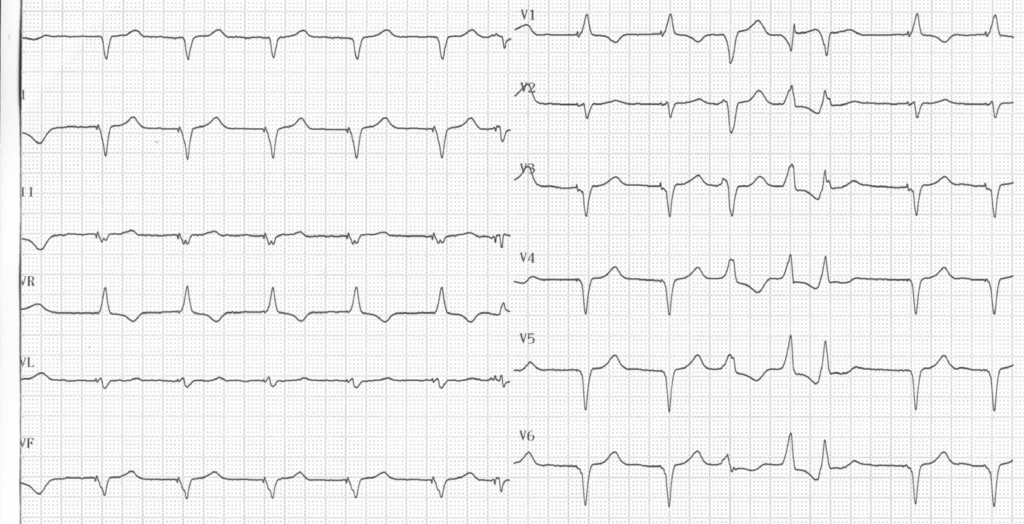
ECG réalisé après la cessation de la tachycardie, montrant une fibrillation auriculaire de faible amplitude, une stimulation biventriculaire (complexe QRS étroit, négatif en dérivation I et positif en V1) ; présence d'un triplet ventriculaire polymorphe attestant d'un contexte arythmogène ;
À retenir
La réentrée inter-branches survient de manière préférentielle chez les patients atteints d'insuffisance cardiaque présentant un bloc de branche gauche associé à une tachycardie fréquente.