


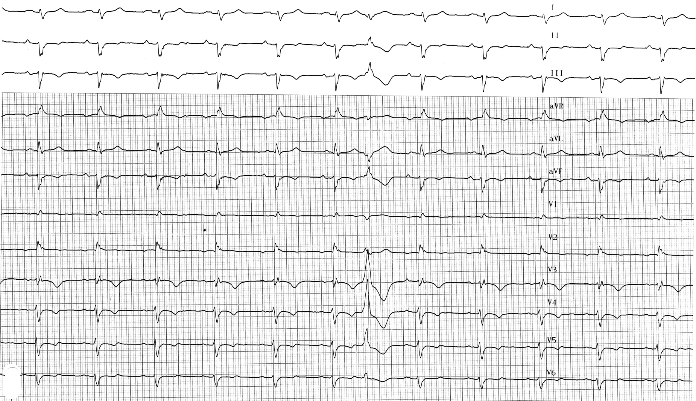
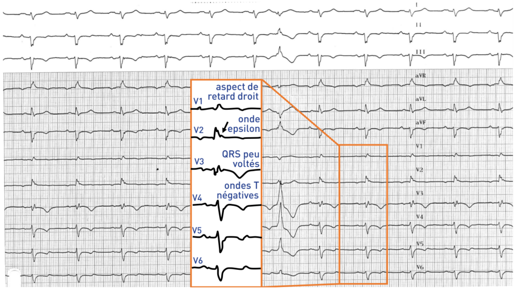
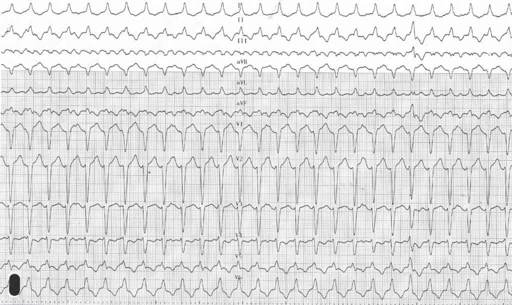
Description du tracé
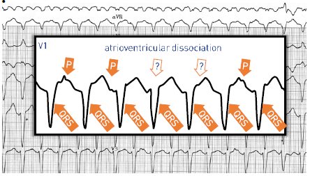
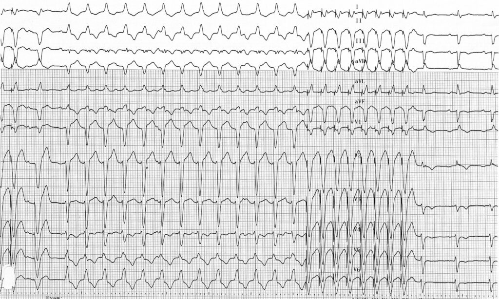
tachycardie ventriculaire relativement lente (130 bpm) avec retard gauche ; transition QRS (R/S > 1) en V5 (tardive) ; dissociation auriculo-ventriculaire confirmant le diagnostic de tachycardie ventriculaire (activité auriculaire clairement identifiable en V1) ; ce tracé correspond à une tachycardie de la voie d'éjection ventriculaire droite (infundibulum pulmonaire) ;
Commentaire
La dysplasie ventriculaire droite arythmogène est une maladie héréditaire caractérisée par le remplacement progressif des cellules du myocarde par du tissu fibro-adipeux. La transmission est autosomique dominante, avec une pénétrance et une expression variables, due à une mutation des gènes codant pour les protéines desmosomiques. La dysplasie touche principalement les hommes, avec un rapport hommes/femmes de 3 pour 1, et se manifeste généralement chez des sujets jeunes, les symptômes apparaissant le plus souvent avant l’âge de 40 ans. Le remplacement du myocarde par du tissu adipeux et une fibrose peut être étendu et s’étendre au ventricule gauche dans les formes avancées, ou être plus localisé au niveau de l’apex ou de l’infundibulum ventriculaire droit. L’évolution de la maladie varie d’un individu à l’autre, certains patients restant totalement asymptomatiques ou ne présentant que des symptômes mineurs. Les anomalies histologiques peuvent contribuer au développement de troubles du rythme ventriculaire pouvant entraîner des palpitations, une syncope ou une mort subite lors d’une activité sportive intense. Cette maladie est fréquemment observée chez les sportifs de haut niveau ayant subi une mort subite et constitue la deuxième cause (après la cardiomyopathie hypertrophique) de mort subite chez le jeune patient. L’une des difficultés dans la prise en charge de ces patients réside dans le fait que la première manifestation clinique observée peut être la survenue d’une mort subite. À mesure que la maladie évolue, des symptômes liés à une insuffisance cardiaque droite, puis biventriculaire, peuvent apparaître.
Le diagnostic repose sur la mise en évidence d'une combinaison de critères cliniques, électrocardiographiques ou morphologiques, tant majeurs que mineurs. L'évaluation a pour objectif de confirmer le diagnostic, d'évaluer le risque de mort subite et de déterminer l'étendue de la maladie.
L'électrocardiogramme de surface joue un rôle central, car certains critères sont considérés comme majeurs et d'autres comme mineurs. Il peut mettre en évidence :
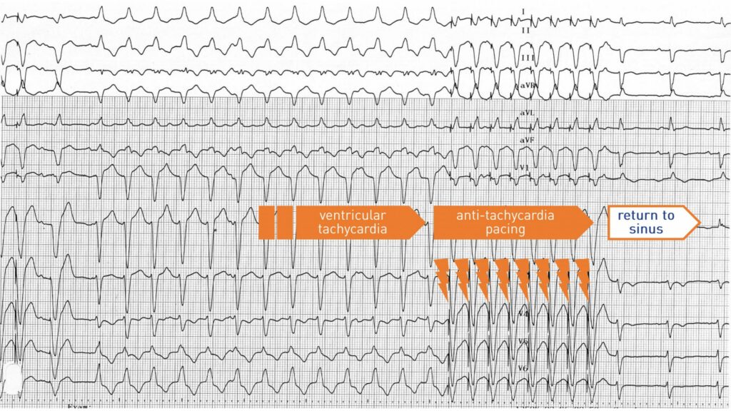
Description du tracé
Ce tracé illustre une séquence de stimulation anti-tachycardique délivrée par le défibrillateur implantable, qui permet d'interrompre la tachycardie et de rétablir le rythme sinusal ;
Commentaire
Outre l'électrocardiogramme, plusieurs examens complémentaires sont réalisés :
La prise en charge thérapeutique varie en fonction de la gravité de la maladie. En cas d’arythmies cardiaques sévères survenant le plus souvent à l’effort, la pratique des sports de compétition est contre-indiquée. Les bêtabloquants à forte dose constituent le traitement médicamenteux de référence. L'ablation par radiofréquence peut être proposée essentiellement chez les patients présentant des épisodes de tachycardie ventriculaire récurrente résistants au traitement médical. Les patients les plus exposés au risque de mort subite doivent bénéficier de la pose d'un défibrillateur. Les facteurs de risque reconnus sont les antécédents de mort subite, les syncopes, les antécédents familiaux de mort subite ou les troubles du rythme récurrents malgré un traitement médical bien contrôlé.
English English French