


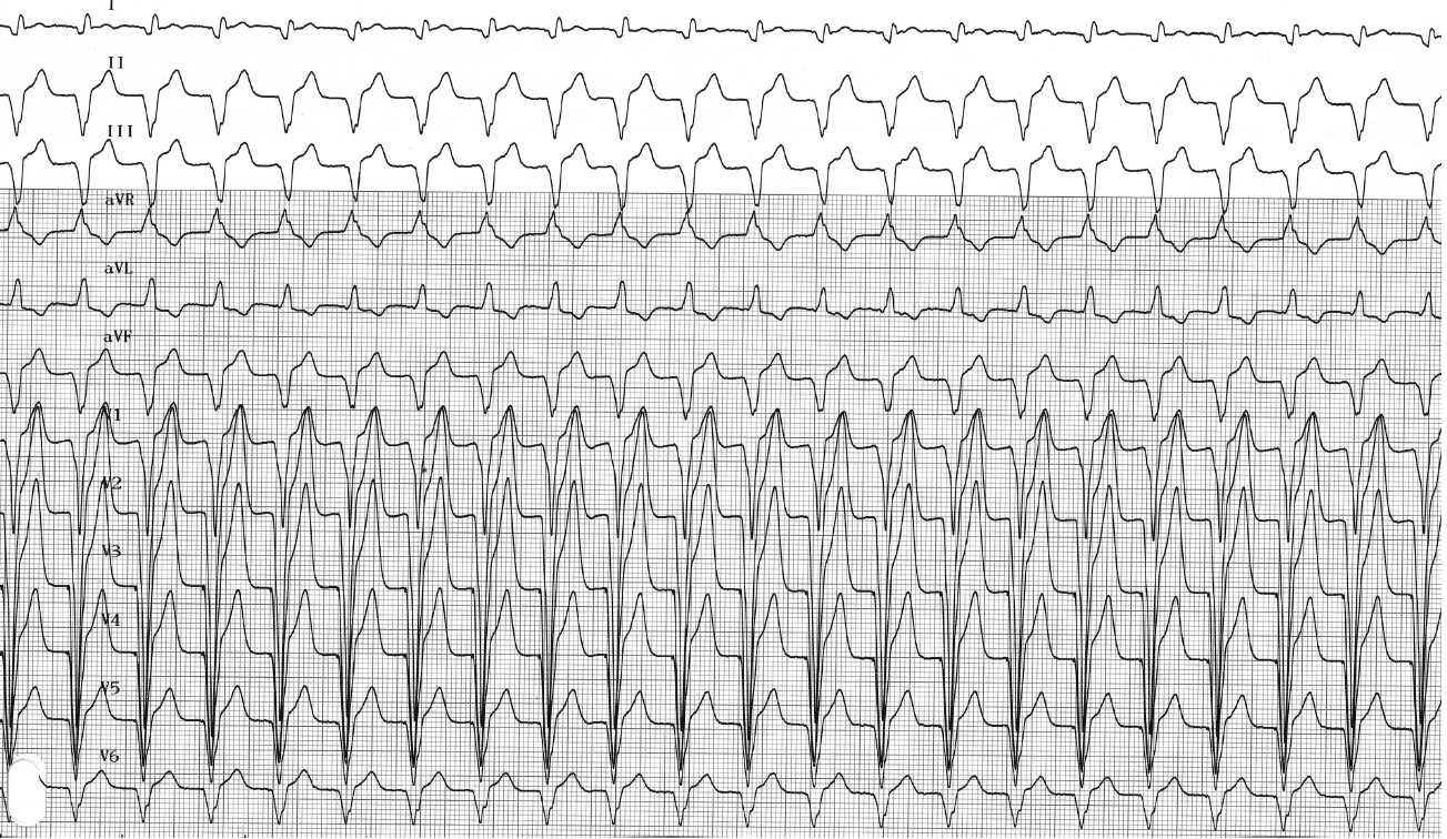
La tachycardie induite par le stimulateur cardiaque (TRE) est le terme utilisé pour décrire une tachycardie d’origine électrique qui survient le plus souvent à la fréquence de stimulation maximale, avec des séquences répétées de stimulation ventriculaire suivies d’une conduction auriculaire rétrograde. Le déclenchement d’un TRE résulte de la détection, en dehors des périodes réfractaires, d’une onde P’ rétrograde, qui provoque un délai AV — souvent prolongé — qui, à son tour, favorise la conduction rétrograde après une stimulation ventriculaire. Un TRE est donc une séquence répétitive dans laquelle le stimulateur cardiaque répond à chaque onde P’ rétrograde en stimulant le ventricule à une fréquence élevée, ce qui génère à son tour une onde P’ rétrograde. Le cycle se répète ainsi indéfiniment, à moins qu’un bloc rétrograde ne survienne ou qu’un algorithme spécifique du stimulateur cardiaque n’intervienne.
La présence d’une conduction rétrograde est donc une condition préalable essentielle au déclenchement et au maintien d’un TRE. La conduction rétrograde est présente chez 40% de patients en moyenne bénéficiant d’une stimulation au repos, toutes indications confondues. Elle est beaucoup plus fréquente chez les patients ayant reçu un implant pour un dysfonctionnement du nœud sinusal que pour un bloc auriculo-ventriculaire. Ces chiffres sont suffisamment élevés pour justifier la programmation systématique d’une protection efficace. Outre la présence d’une conduction rétrograde, le déclenchement d’un TRE nécessite la programmation d’un mode de stimulation auriculaire (DDD ou VDD) et entraîne une perte temporaire ou permanente de la synchronisation auriculo-ventriculaire. En effet, si l’activité ventriculaire est correctement synchronisée avec celle de l’oreillette, la conduction rétrograde est bloquée. Les événements suivants peuvent favoriser une dissociation auriculo-ventriculaire temporaire, une conduction rétrograde et le déclenchement d’un TRE : une extrasystole ventriculaire (la cause la plus fréquente), un battement prématuré auriculaire accompagné d’un allongement du délai AV afin de maintenir la fréquence cardiaque maximale programmée, un délai AV programmé trop long (le faisceau auriculo-ventriculaire est sorti de sa période réfractaire au moment de la stimulation ventriculaire), des interférences externes ou des myopotentiels détectés par la sonde atriale, un échec de la détection ou de la stimulation auriculaire…
Les premiers stimulateurs cardiaques n'étaient pas protégés contre ce type d'activité incontrôlée, et la tachycardie pouvait persister indéfiniment, provoquant des symptômes parfois très invalidants. Un TRE prolongé peut en effet être difficile à supporter, les symptômes allant d'une simple sensation d'inconfort ou de palpitations à une décompensation cardiaque chez les patients atteints d'une cardiopathie sous-jacente.
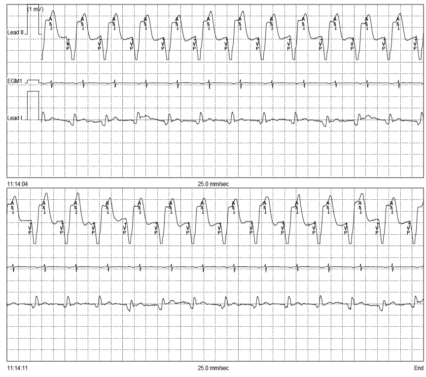
Description du tracé
L'analyse de l'EGM confirme le diagnostic de tachycardie par réentrée électrique : une séquence ininterrompue de cycles VP-AS (stimulation ventriculaire, détection auriculaire) avec un délai AV prolongé afin de garantir que la fréquence maximale synchrone ne soit pas dépassée ;
English English English French English English English French