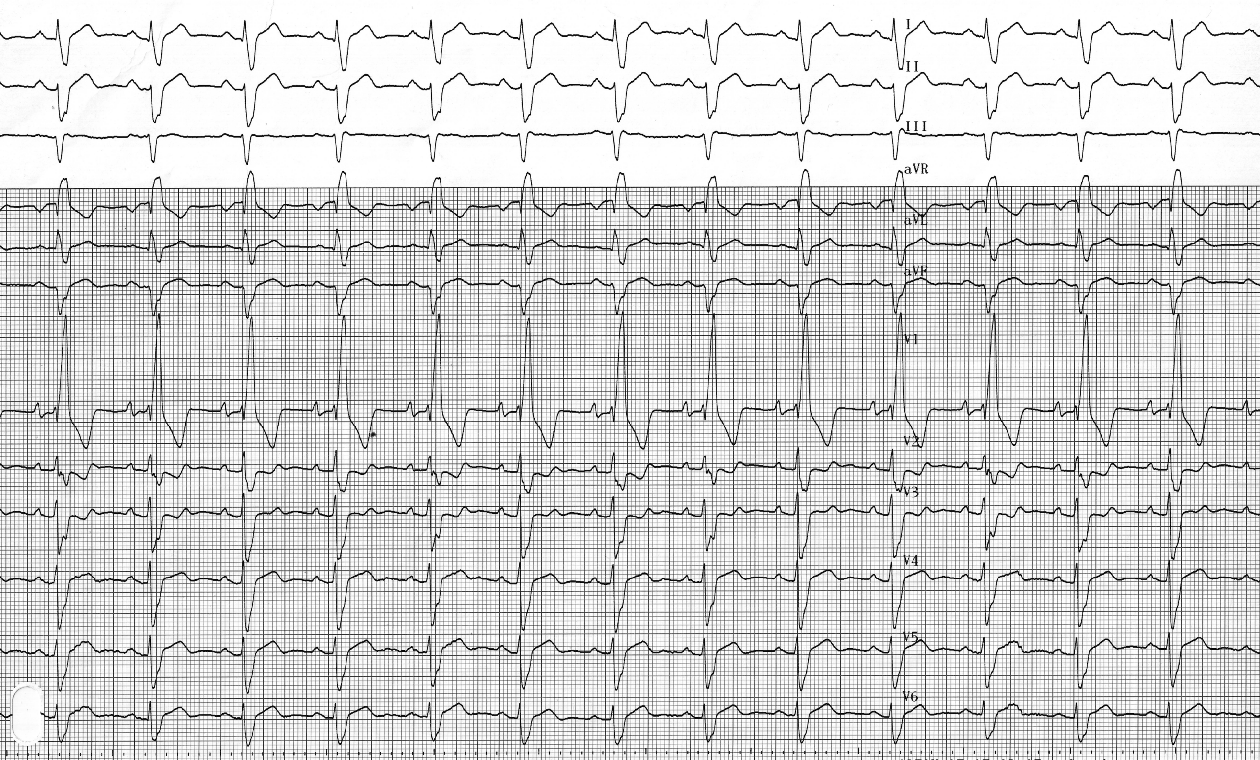
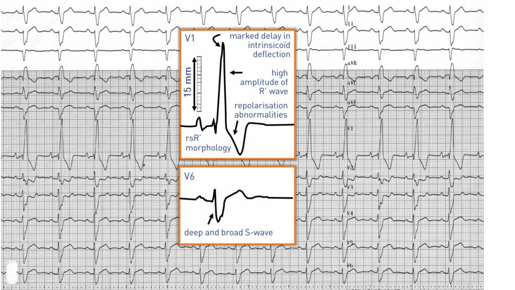
Tracé 3.6 : Bloc de branche droite et hypertrophie ventriculaire droite
Informations destinées aux patients
Homme de 64 ans présentant un cœur pulmonaire chronique (antécédents d'embolie pulmonaire)
ECG
Question
Quel(s) diagnostic(s) correspond(ent) à cet ECG ?
Commentaires
Description de l'ECG
Activité sinusale auriculaire, intervalle PR normal (190 ms), bloc de branche droit complet (durée du QRS de 180 ms, tracé rsR’ en V1) avec hypertrophie ventriculaire droite (onde R’ de forte amplitude en V1, onde S très profonde et large dans les dérivations I, V5, V6, déviation intrinsèque nettement retardée en V1, troubles de la repolarisation de V1 à V3) ;
Explication
Le diagnostic électrocardiographique de l'hypertrophie ventriculaire droite est difficile en présence d'un bloc de branche droit et ne se manifeste que si l'hypertrophie est anatomiquement significative.
En cas de bloc de branche droit complet, les signes suivants sont caractéristiques d'une hypertrophie :
• Une onde R’ de forte amplitude, supérieure à 15 mm, dans la dérivation V1
• Une onde S très large dans la dérivation I
• Un retard important de la déviation intrinsèque dans les dérivations précordiales droites
Ces différents paramètres sont observés chez ce patient ; l'hypertrophie ventriculaire droite est en effet fréquente chez les patients atteints de cœur pulmonaire chronique.
À retenir
Une déviation importante de l'axe vers la droite chez un patient présentant un bloc de branche droit peut refléter la présence d'une hypertrophie ventriculaire droite ou d'un bloc fasciculaire postérieur gauche associé. La présence d'une onde R’ de forte amplitude est particulièrement évocatrice d'une hypertrophie ventriculaire droite.