


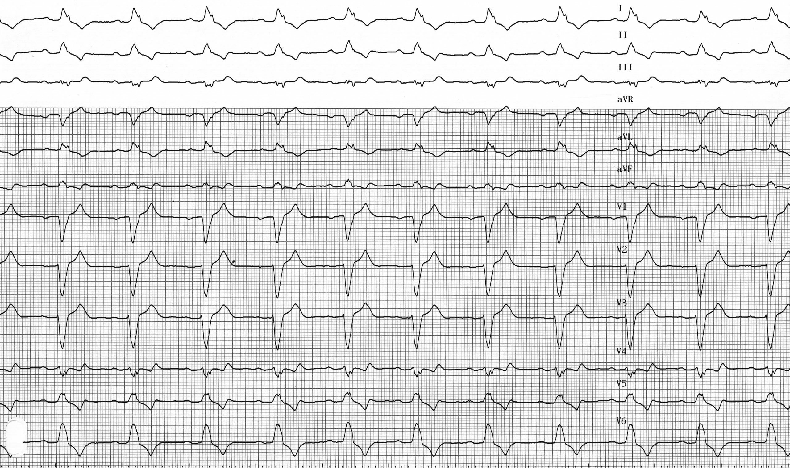
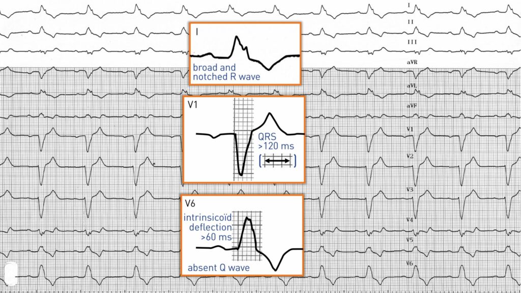
Ce patient présente un tracé électrocardiographique caractéristique d'un bloc de branche gauche, associant :
1. Un complexe QRS élargi de 120 ms ou plus chez l'adulte, et de plus de 100 ms chez les enfants de moins de 8 ans ;
2. Un retard de l'apparition de la déviation intrinsicoïde supérieur à 60 ms (temps d'activation ventriculaire pouvant atteindre jusqu'à 100 ms) dans les sondes ventriculaires gauches : cela est dû au retard de dépolarisation de la paroi ventriculaire touchée ; il s'agit d'un critère majeur et fondamental du bloc de branche gauche ; la déviation intrinsèque est normale dans les dérivations précordiales droites, car le ventricule droit est activé normalement ;
3. Un tracé QRS caractéristique en V6 : on observe une déviation initiale positive, la direction de la dépolarisation septale étant inversée et se propageant de droite à gauche ; les vecteurs d’activation septale sont ainsi dirigés vers les dérivations précordiales gauches qui enregistrent une déviation initiale positive ; la présence d’une onde q en V6 permet donc d’écarter le diagnostic de bloc de branche gauche ; l’amplitude de l’onde R est souvent accrue ; en effet, lorsque l’activation ventriculaire gauche est retardée, elle ne rencontre plus d’opposition de la part de la dépolarisation ventriculaire droite, ce qui explique un potentiel de tension plus élevé ; le pied de l’onde R est souvent flou ou parfois entaillé, et la branche ascendante qui suit est relativement rapide, puis suivie d’un ralentissement considérable au sommet (bifidité ou profil en plateau) et d’une brève descente vers la ligne isoélectrique ; absence d’onde S terminale ;
4. Un tracé QRS caractéristique en V1 : une onde S large associée à une apparition retardée des vecteurs de dépolarisation gauche ; le tracé le plus typique est donc rS ou QS ;
5. Modification du segment ST et de l'onde T dans les dérivations V5 et V6 : on observe fréquemment un affaissement du segment ST et une inversion de l'onde T, pouvant s'accompagner d'une élévation dans la dérivation V1 ;
6. Dans les dérivations frontales, le tracé caractéristique observé en V6 (positivité exclusive suivie d'une onde T négative et symétrique avec dépression) se retrouve en dérivation I et, plus souvent encore, en aVL ;
L'axe QRS est variable : il peut être normal ou dévié vers la gauche. La présence d'un bloc de branche gauche complique le diagnostic d'infarctus ou d'hypertrophie ventriculaire.
English English English French English English English French