BiV markers with changing far-field morphology
Patient
Go to the casebook version optimized for the mobile experience
RESONATE X4 CRT-D implanted 4½ years earlier, sinus rhythm; DDD 50-130 bpm, sensed AV delay 120 ms and paced AV delay 150 ms, both fixed (programmed 120-120 and 150-150 ms), BiV with LV offset 0 ms
Remote transmission, no alert; counters since a reset 3 months after implant: RV and LV paced 98%, atrial pacing 3%, 14.8 million PACs counted (about 9,300 a day)
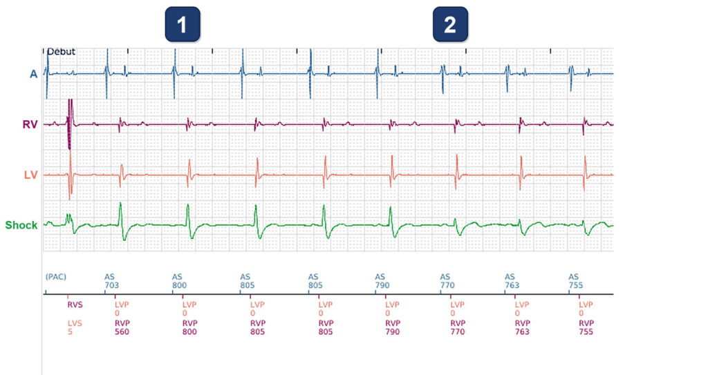
Trace
- Real-time EGM of the transmission (channels A, RV, LV, Shock): sinus rhythm at about 75 bpm, each atrial event tracked; LV and RV paced together 120 ms after it (AS 800 → LVP 0, RVP 800: sensed AV delay 120 ms, LV offset 0 ms). On the shock EGM, a tall narrow spike then a deep negative wave: the fully paced complex
- From here on (AS 770, 763, 755 ms) the same markers, LVP and RVP 120 ms after each atrial event, but another shock EGM: a small notched complex, no tall spike. The pulses meet the patient’s own activation already under way: fusion, still marked and counted as BiV
- An atrial event conducts before the 120 ms sensed AV delay ends: the RV is sensed about 110 ms after it (AS 740 → RVS 733), the LV 13 ms later (LVS 13), and no pulse is delivered. The shock EGM of this conducted beat, like that of the first beat of the first strip (RVS, LVS 5), is much like the fused beats
- PAC 383 ms after that atrial event: pacing 120 ms after it would exceed the maximum tracking rate (130 bpm = 460 ms), so the pace is held back. The PAC conducts first: the RV is sensed about 185 ms after it (RVS 455), just before the 460 ms limit
- After the PAC the paced beats capture fully again: tall spike and deep negative wave on the shock EGM (AS 695 → LVP 0, RVP 630), and so on the next 4 beats. The same is seen after the two other PACs of this 30-s recording
- Sixth beat after the PAC (AS 790): the spike shrinks again, fusion is coming back. With the patient’s own AV conduction about as fast as the sensed AV delay, every beat is a race between the pulses and the conducted activation
- Heart rate over 6 months, one value a day (max, mean, min; vertical lines = the 1st of each month): mean 69-78 bpm until just before the first line, about 75-90 bpm over the next 4½ months, 93-110 bpm for 10 days, 2 to 3 weeks before the transmission, then 80 bpm the day before it
- % paced, RV and LV curves on top of each other (atrial pacing near 0%): 98-99% until just before the first line, mostly 90-97% over the next 4½ months, 64-77% during the 10 days of fastest rate, then 93% RV and 92% LV the day before the transmission. Fused beats count as paced: these figures overstate the CRT delivered
Comments
BiV markers on nearly every beat, yet CRT is partly lost: the changing shock (far-field) EGM shows fusion with the patient’s own AV conduction, about as fast as the 120 ms sensed AV delay. Some beats are fully conducted, after a sinus atrial event and after a PAC. The sensed AV delay needs to be shortened, below the patient’s own AV conduction
Points to remember
- Compare the beats on the far-field (shock) EGM: the fully paced beat keeps one shape; a smaller or notched complex under the same LVP and RVP markers is fusion with the patient’s own conduction
- Fusion comes when the patient’s AV conduction is about as fast as the sensed AV delay (here about 110 ms against a fixed 120 ms). When a sinus atrial event or a PAC conducts first, the beat is not paced at all (RVS, LVS)
- The % paced counts every LVP and RVP, fused or not: 92-98% here overstates the CRT delivered. The trend shows its link with the heart rate: 64-77% paced when the mean rate was 93-110 bpm
- The sensed AV delay needs to be shortened: to keep the pacing ahead of the patient’s own activation, it must be clearly shorter than the patient’s AV conduction (here it is longer: 120 ms against about 110 ms); after any change, check the shock EGM shape again
Take home message
A BiV marker is not BiV capture: fused beats carry the same LVP and RVP markers and count as paced, so 98% BiV pacing can hide a partial loss of CRT. The far-field (shock) EGM shows the difference. Here the sensed AV delay needs to be shortened, so that the pacing comes before the patient’s own conduction
Read more about related topics