


Description du tracé
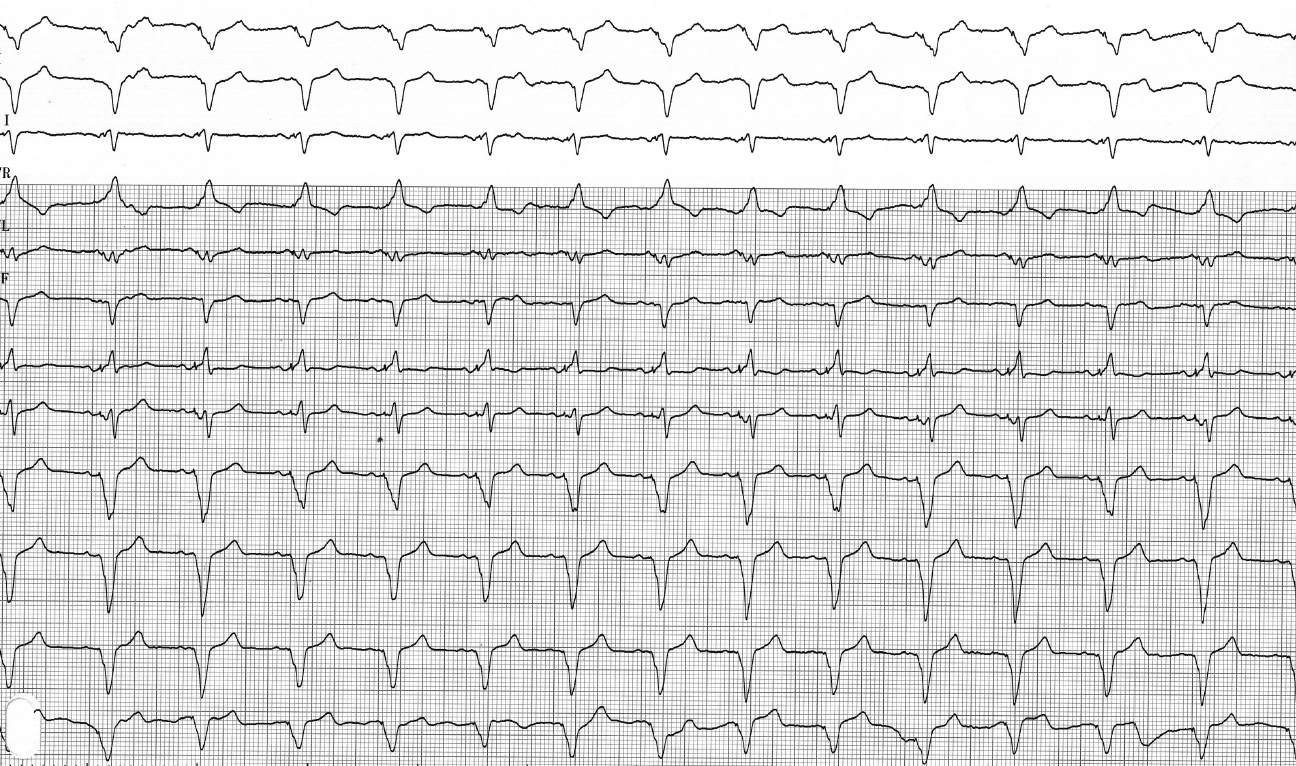
Enregistrement ECG réalisé après programmation d'une stimulation ventriculaire gauche uniquement ; le complexe QRS est nettement plus large que lors d'une stimulation biventriculaire, avec une négativité marquée en DI et une positivité en V1, ce qui correspond à la position latérale de la dérivation ;
Commentaire
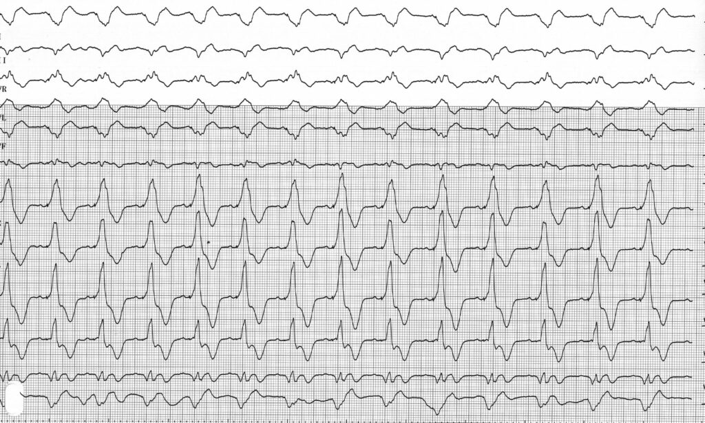
L'électrocardiogramme est un outil essentiel dans le suivi des patients bénéficiant d'une thérapie de resynchronisation cardiaque ; il peut faire suspecter un déplacement entraînant une perte de capture ventriculaire gauche, même si le diagnostic définitif repose sur l'interrogation du stimulateur cardiaque et une radiographie thoracique. Afin de faciliter le suivi par le cardiologue référent, il est utile de disposer d’un électrocardiogramme de référence du patient montrant le rythme spontané, la capture ventriculaire droite seule, la capture ventriculaire gauche seule et la stimulation biventriculaire. Cela permet d’identifier facilement une perte de capture par comparaison. Malgré un remodelage potentiellement important en termes de volume ventriculaire, l’aspect de chaque configuration évolue relativement peu au fil du temps, tant en termes de largeur que de morphologie du complexe QRS.
Pour tenter de diagnostiquer une perte de capture sur l'électrocardiogramme, il est nécessaire de bien comprendre certains facteurs. Le schéma de stimulation biventriculaire dépend de la fusion de deux champs d'activation distincts provenant respectivement de la sonde ventriculaire droite et de la sonde ventriculaire gauche. Le schéma observé lors de la stimulation biventriculaire varie d’un patient à l’autre en fonction de la position des deux sondes de stimulation, de la partie du myocarde dépolarisée respectivement par la sonde droite et la sonde gauche, des caractéristiques électriques et anatomiques des patients, ainsi que du degré de fusion avec l’activation spontanée.
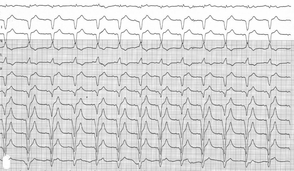
La pathologie la plus courante et la plus importante sur le plan clinique à diagnostiquer est la perte de capture ventriculaire gauche. La durée du QRS est généralement plus courte lors d’une capture biventriculaire que lors d’une stimulation monoventriculaire droite ou gauche, mais ce critère à lui seul n’est pas suffisamment discriminant pour mettre en évidence une perte de capture. Le diagnostic est facilité lorsque la sonde ventriculaire droite est placée au niveau de l’apex. En effet, la stimulation ventriculaire droite apicale s’accompagne, dans la majorité des cas, d’un QRS positif en DI et d’un QRS négatif dans les dérivations inférieures. L’axe est généralement dévié vers la gauche, et le QRS n’est presque jamais positif en V1. Il convient de rechercher un QRS négatif en DI et/ou un QRS positif en V1.
En effet, la présence de l’un ou l’autre de ces deux signes (une onde négative en DI et/ou une onde positive en V1) confirme l’existence d’une capture ventriculaire gauche effective. Un tracé compatible avec une stimulation monoventriculaire droite n'exclut toutefois pas l'existence d'une capture ventriculaire gauche efficace, mais peut être lié à l'influence prédominante de la sonde droite sur le complexe QRS. La durée du QRS est souvent plus courte lors d’une stimulation biventriculaire que dans le profil observé lors d’une stimulation ventriculaire droite. Lorsque la sonde ventriculaire droite est située dans le septum supérieur ou au niveau de l’infundibulum, le diagnostic d’une perte de capture ventriculaire gauche est beaucoup plus difficile, car le profil observé en V1 et DI n’est plus aussi caractéristique.
La perte de capture ventriculaire droite est moins fréquente, mais moins préjudiciable sur le plan hémodynamique. Le complexe QRS lors de la stimulation ventriculaire gauche est souvent nettement élargi. L’axe du QRS est droit ou hyper-droit. Sa morphologie dépend de la position de la sonde ventriculaire gauche. Plus la sonde est latérale, plus le QRS devient négatif dans la dérivation DI, avec un aspect de bloc de branche droit. Plus la sonde est apicale, plus le QRS devient négatif dans les dérivations inférieures.
Description du tracé
Tracé enregistré après programmation d'une stimulation ventriculaire droite seule ; le complexe QRS est plus large que lors d'une stimulation biventriculaire, avec une négativité marquée dans les dérivations inférieures, compatible avec la position apicale de la sonde de stimulation ;
English English French