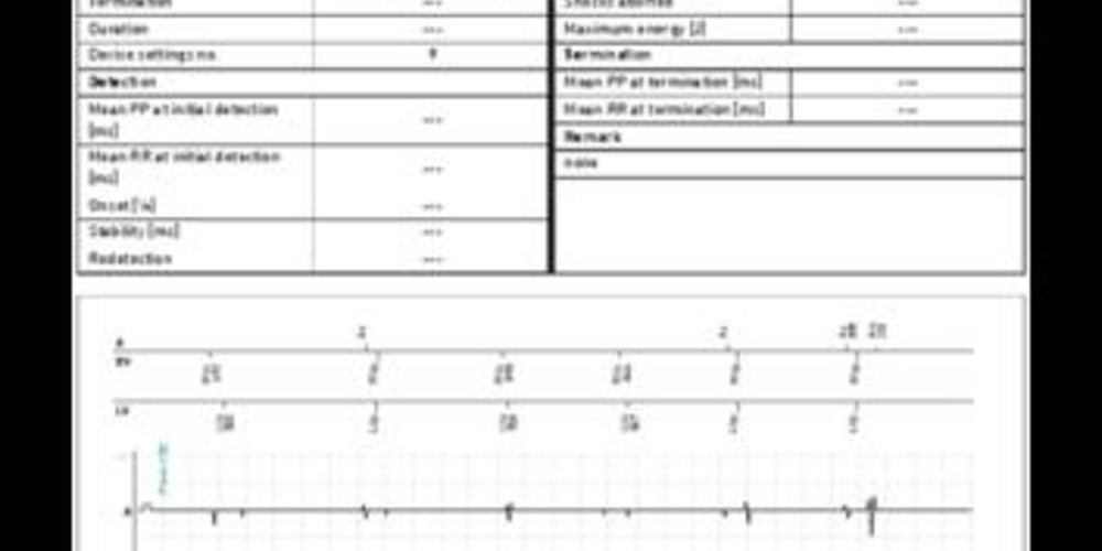
double counting of the R wave from ventricular extrasystole
Tracing
Manufacturer Biotronik
Device Remote monitoring
Field Periodic Egm
N° 3
Patient
This 54-year-old man underwent implantation of a Lumax 540 HF-T triple chamber defibrillator in the context of ischemic cardiomyopathy with left bundle branch block; periodical EGM;

Graph and trace
Telecardiology tracing
The 4 available channels are the markers with the time intervals, the atrial sensing channel, and the right and left ventricular sensing channels, each for 30 seconds.
- sinus rhythm with biventricular stimulation;
- ventricular pair;
- VES, probably originating from the right ventricle, sensed first in the right then in the left ventricular channel;
- VES with oversensing in the right ventricular channel of a supernumerary ventricular electrogram, corresponding to the double counting of the R wave at the end of the ventricular blanking period;
- further VES oversensing.
Other articles that may be of interest to you


This patient had to be seen in a face-to-face consultation to resolve several issues. The main one was the double counting of the R wave, limited to some VES of right ventricular origin. Should ventricular tachycardia develop, the risks associated with double counting were: 1) erroneous classification of tachycardia at 300 bpm (in the VF zone) instead of 150 bpm; 2) unnecessarily forceful therapies, such as electrical shocks instead of anti-tachycardia pacing. As in the case of the previous patient, the programming of the blanking period (80 ms) was probably too short, and a lengthening of the programmed value to 110 ms resolved the issue.
Ventriculo-atrial crosstalk requires a lengthening of the post-ventricular stimulation atrial blanking period to prevent possible erroneous diagnoses of atrial arrhythmias with mode switch to DDI pacing, which might cause a loss of biventricular resynchronization as long as the device is in mode switch.
From a clinical perspective, the VES were frequent at the time of the recording of the periodic EGM. A careful surveillance of their occurrence on the successive reports of telecardiology, and an understanding of their clinical consequences was in order. If frequent, a search for a metabolic abnormality and a surveillance of the evolution of left ventricular function were required in this patient suffering from heart failure.