
Chapter 3 · Discrimination · Case 10
Dual-chamber discrimination and dual tachycardia
Patient and episode
Patient
- 59-year-old man; mitral valve cardiomyopathy with reduced ejection fraction; implanted with a dual-chamber defibrillator
Summary
- Episode diagnosed as dual tachycardia by defibrillator with AF and VT
- A>V but stable rhythm and discordant vectors between tachycardia and the reference vector
The recording
Tap a number on the trace, or an entry in the list below
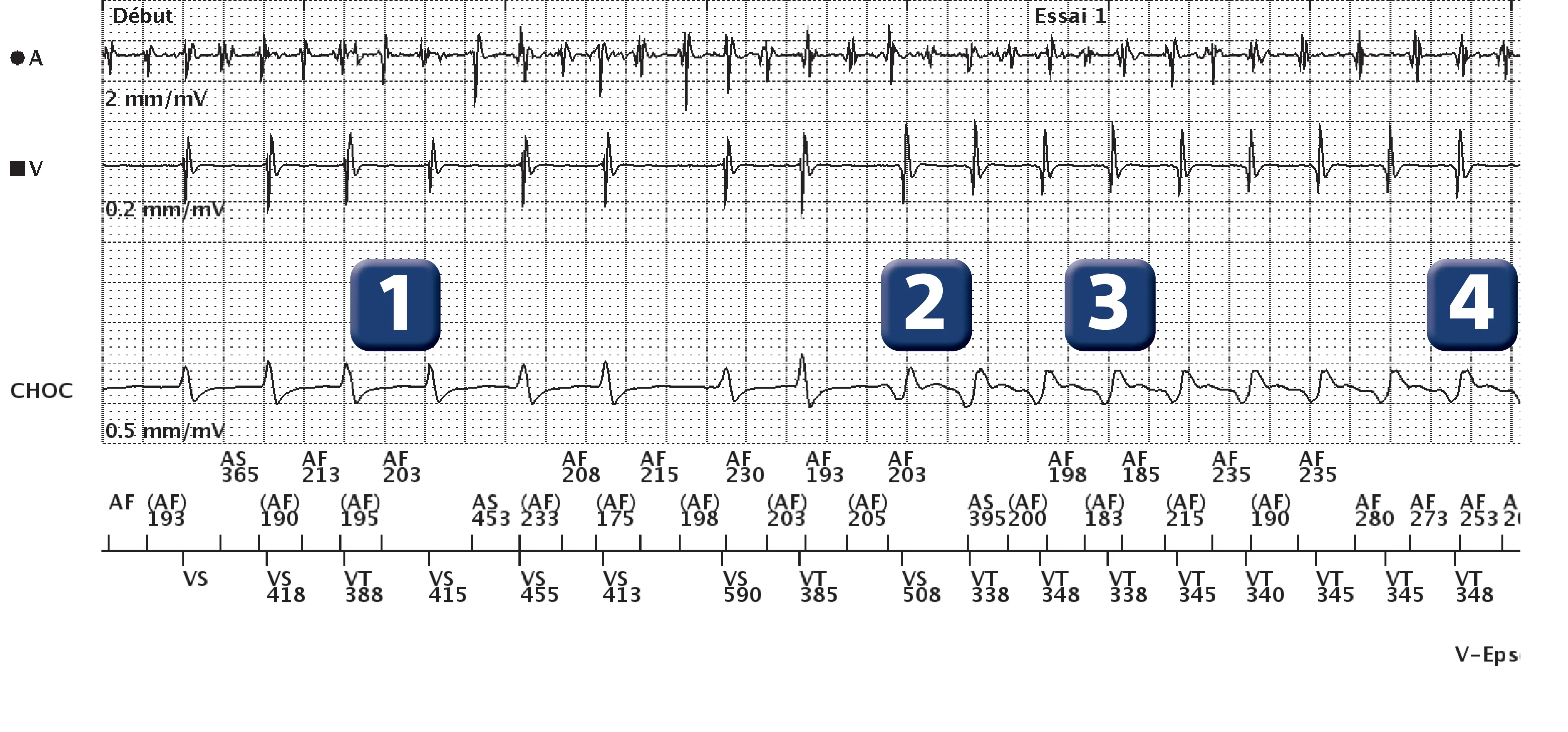
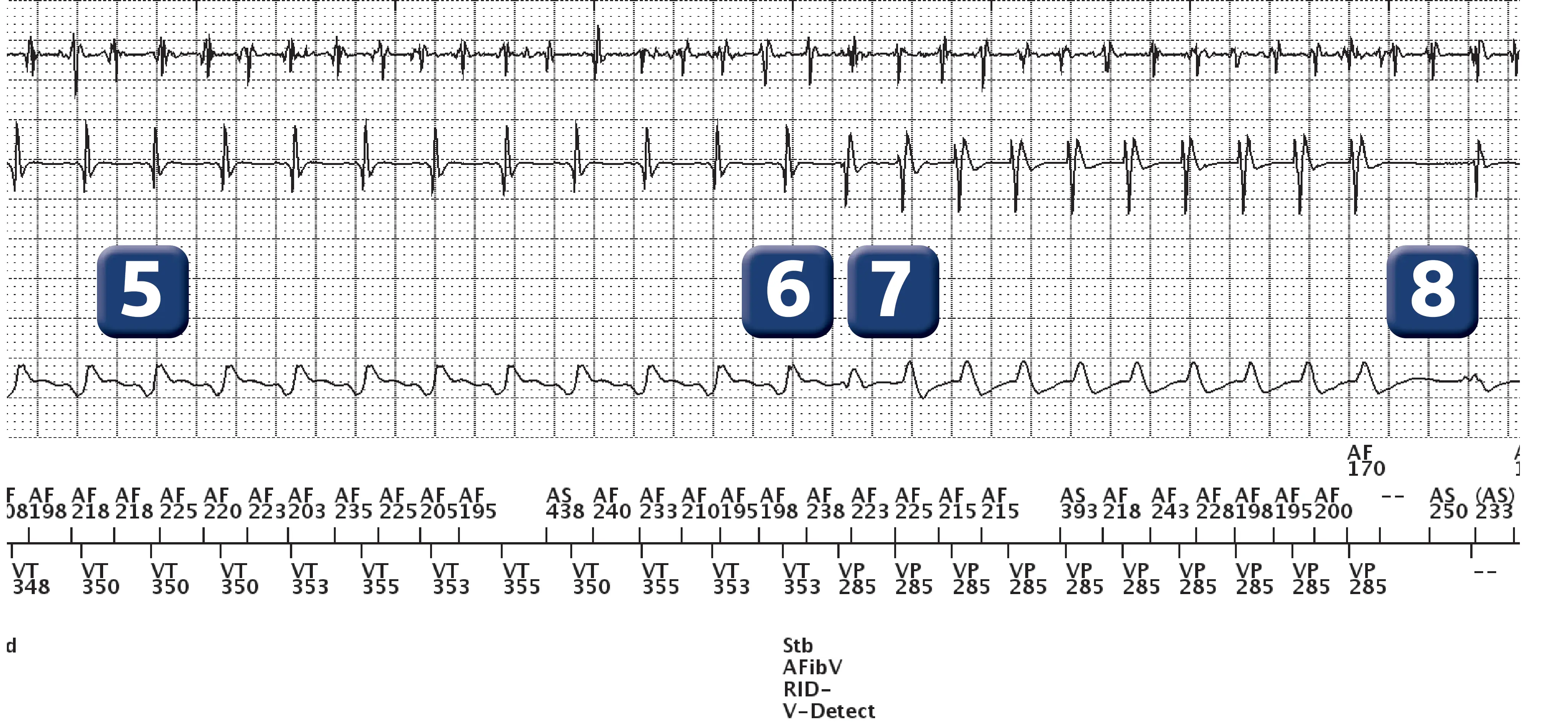
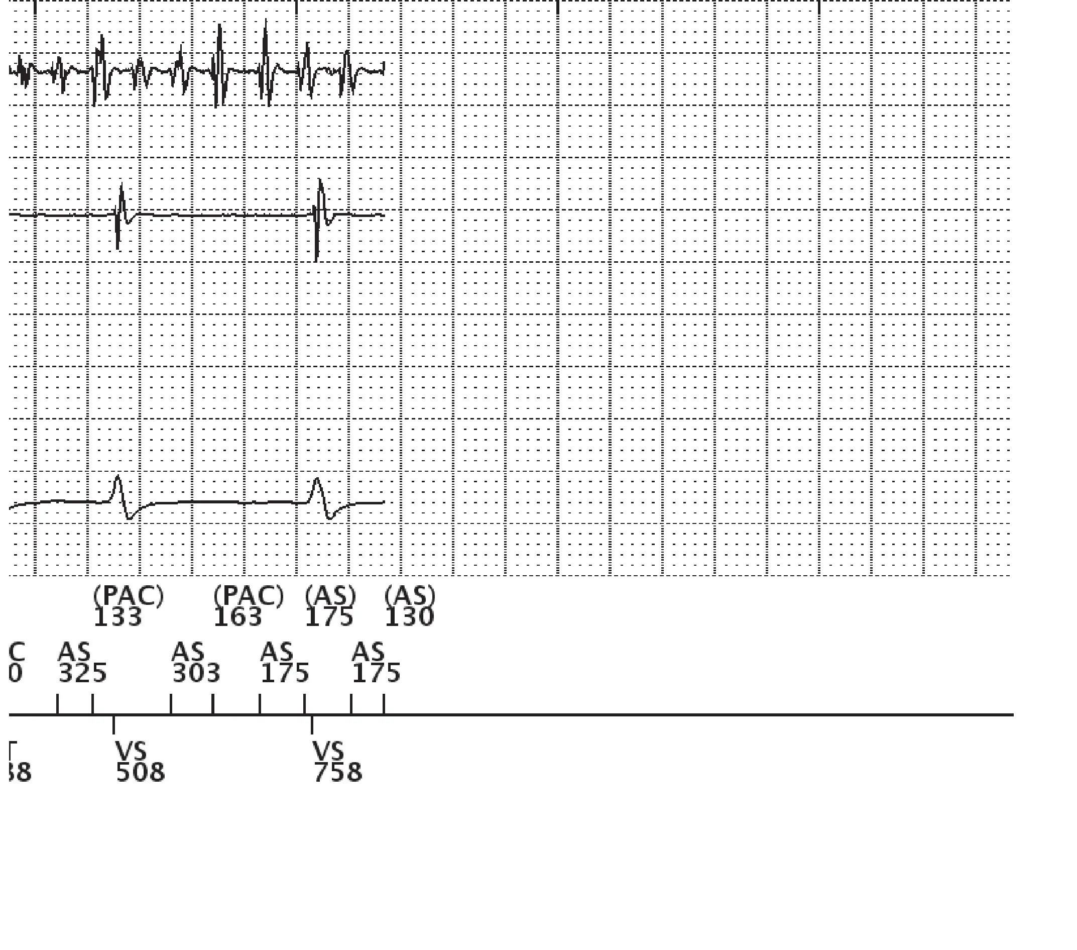
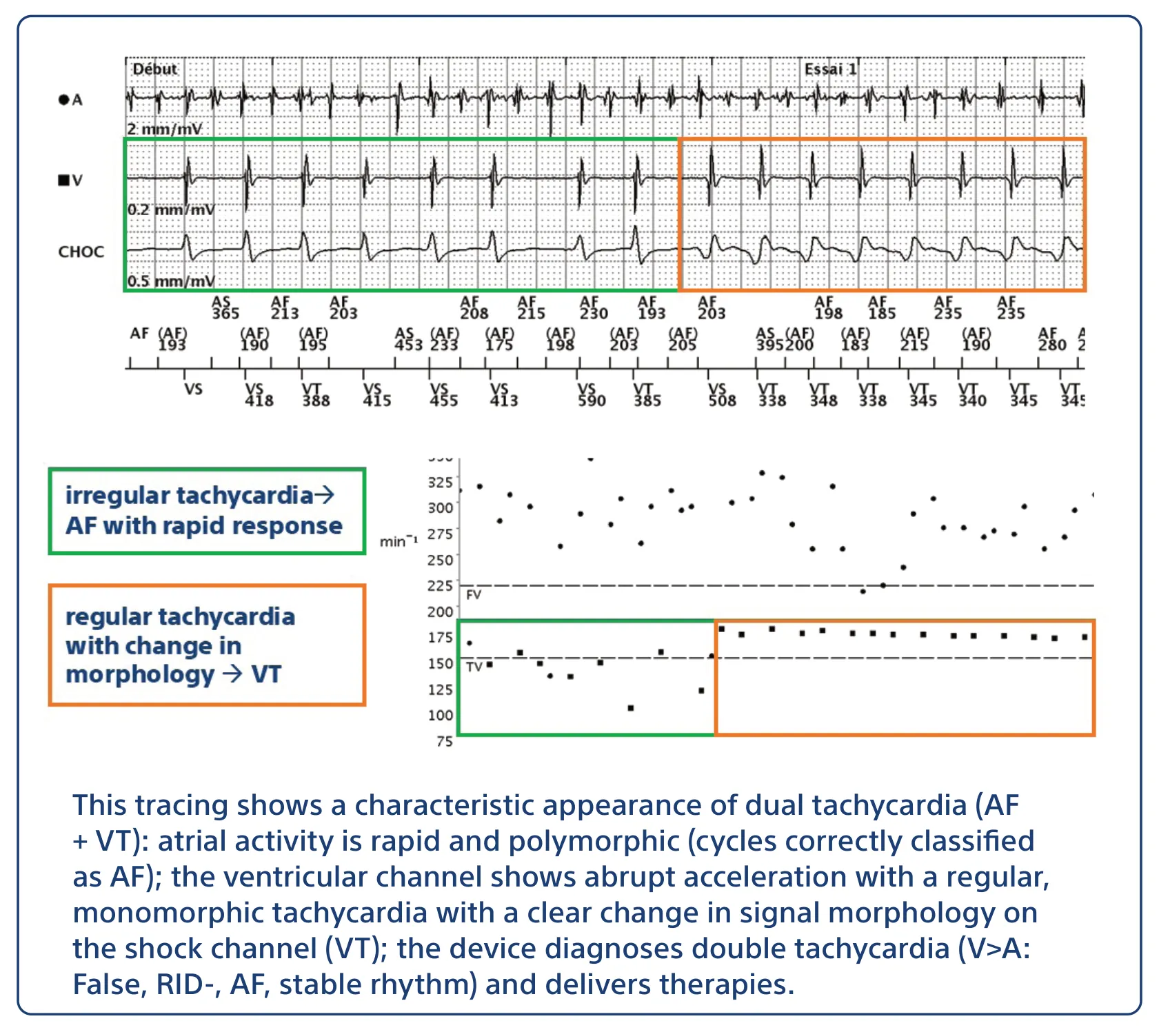
- AF with relatively rapid and irregular atrioventricular conduction
- Sudden acceleration of ventricular rhythm with change in ventricular electrogram morphology; rhythm becomes regular
- At the third consecutive cycle in VT zone, initiation of arrhythmia episode detection; from this interval onwards, each atrial interval is classified as below or above the atrial fibrillation threshold (in this patient, threshold set at 170 beats/minute, nominal value); it is also from this cycle onwards (5 cycles before the episode marker) that stability measurement begins; the mean variance is calculated over these 5 intervals (4 variances)
- After 8 consecutive VT cycles, V-Epsd marker
- From this cycle, 10 cycles remain until the V-Detect marker is reached; vector correlation analysis is performed over these 10 cycles.
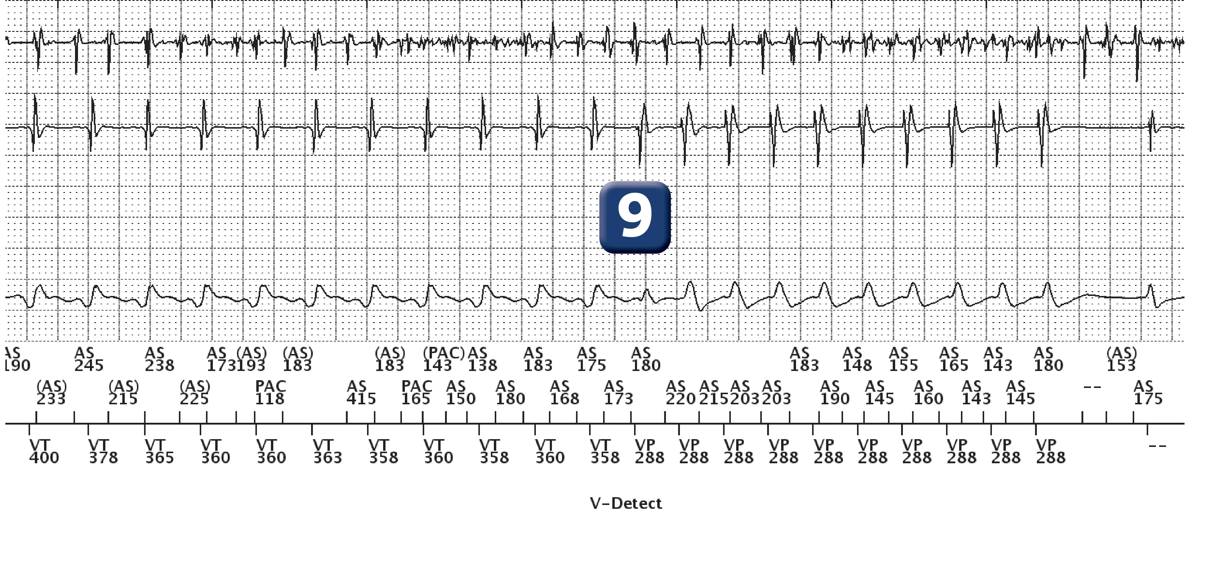
- At the end of the duration, diagnosis of dual tachycardia and decision to treat: AF (initially at least 6 out of 10 rapid atrial intervals, then at least 4/10 rapid intervals in a rolling window), stable rhythm (measured variability < programmed stability threshold) without correlation between morphology in tachycardia and reference morphology (for less than 3 beats/10, the vector correlates with the reference vector).
- ATP burst (burst of 10 complexes)
- Probable burst failure and ongoing dual tachycardia
- Confirmation of persistent tachycardia based solely on a heart rate criteria (V-Detect). No discrimination is applied post-ATP
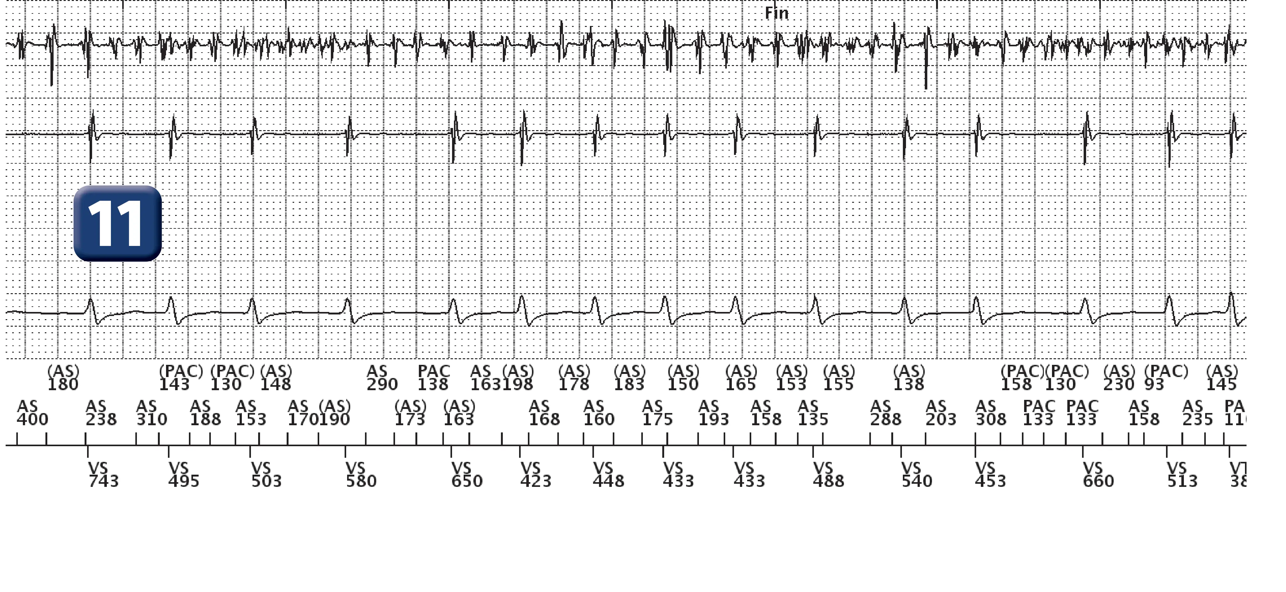
- Second burst
- Effective burst and termination of ventricular arrhythmia
Points to remember
- This trace shows the value of coupling discriminators for the diagnosis of complex tachycardias such as dual tachycardias; discrimination is performed in stages
- Stage 1, V>A : False
- Stage 2, at least 3 ventricular cycles out of 10 correlated: false (RID-)
- The analysis continues (step 3) with an analysis of stability and a search for AF
- The AF rate threshold analysis looks for the presence of a fast atrial rhythm, by comparing the various atrial intervals against a programmed threshold (A Fib rate threshold); the atrial analysis begins when ventricular tachyarrhythmia detection is initialized; each atrial interval is classified as either faster or slower than the A Fib rate threshold; when at least 6 of the last 10 intervals are considered to be faster than the A Fib rate threshold, the device declares that AF is present; this parameter can only be programmed in conjunction with stability
- Stability analysis is performed by measuring the degree of variability in RR intervals during tachycardia; the differences between ventricular cycles, as well as an average difference, are calculated throughout the Duration; when the Duration expires, rhythm stability is assessed by comparing the current average difference with the programmed Stability and “Shock if unstable” thresholds; if the average difference is greater than the programmed thresholds, the rhythm is declared unstable; independent thresholds are available for the Stability (inhibitory) and “ Shock if unstable” functions
- The idea behind linking the stability and AFib rate threshold criteria is to treat stable rhythms and unstable rhythms without AF (atrial rate < AFib rate threshold), but to inhibit in the presence of an unstable rhythm with atrial rate > AFib frequency threshold (suspicion of conducted AF).
- In this example, the device finds a diagnosis of AF with a stable rhythm (diagnosis of dual tachycardia) and therapies are delivered
Practice out loud
Just describe the tracing. What do you think is going on? Describing the numbers can help.
00:00
0%
—
From Boston Scientific ICD — clinical cases by P. Bordachar, M. Strik, A. Thiyagarajah, S. Ploux. Published by Cardiocases. Every numbered marker on a recording is explained in the list beneath it; tap a marker on the trace or an entry in the list.
Order hardcopyon Amazon