
Chapter 1 · LV pacing and threshold · Case 4
Non-responder despite 100% BiV pacing
Patient and episode
Patient
- COGNIS CRT-D in a 67-year-old man with dilated cardiomyopathy and left bundle branch block
- No response 6 months after the implant (still breathless on daily activities), although interrogation shows 100% BiV pacing
The recording
Tap a number on the trace, or an entry in the list below
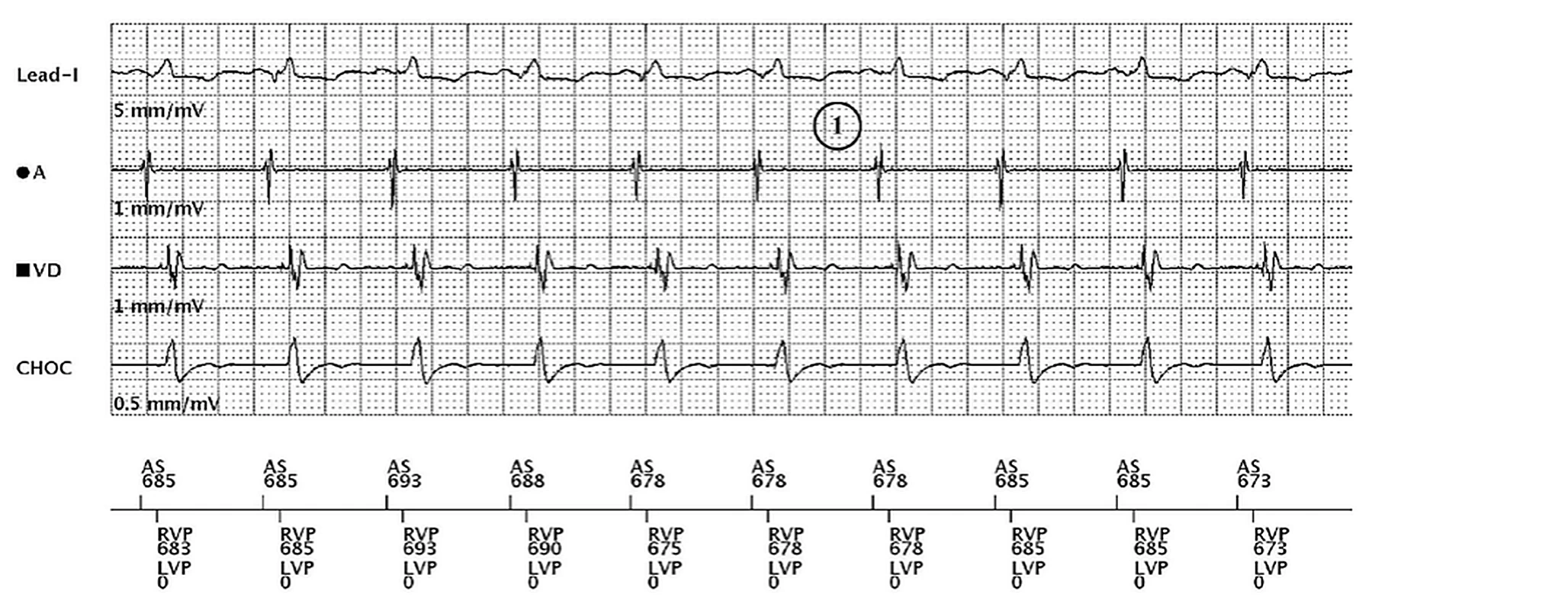
- Recorded at the visit: sinus rhythm (AS ≈ 680 ms), each atrial sensed event tracked by an RV and an LV pace delivered together (RVP, LVP 0: no LV offset), so the counters show 100% BiV pacing; the paced QRS on Lead I is the same on every beat, positive
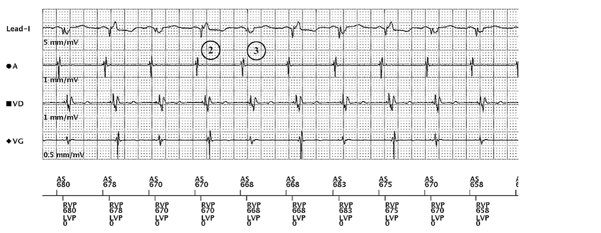
- Same programming, LV output 2.5 V/0.4 ms: this QRS is the same as on the first tracing (positive on Lead I) = RV capture only, the LV is not captured
- The next QRS is different (negative on Lead I) = BiV capture; the two alternate from beat to beat, and the LV EGM (VG) alternates with them. The LV threshold measured at this visit is 2.5 V/0.4 ms: the output sits exactly at threshold
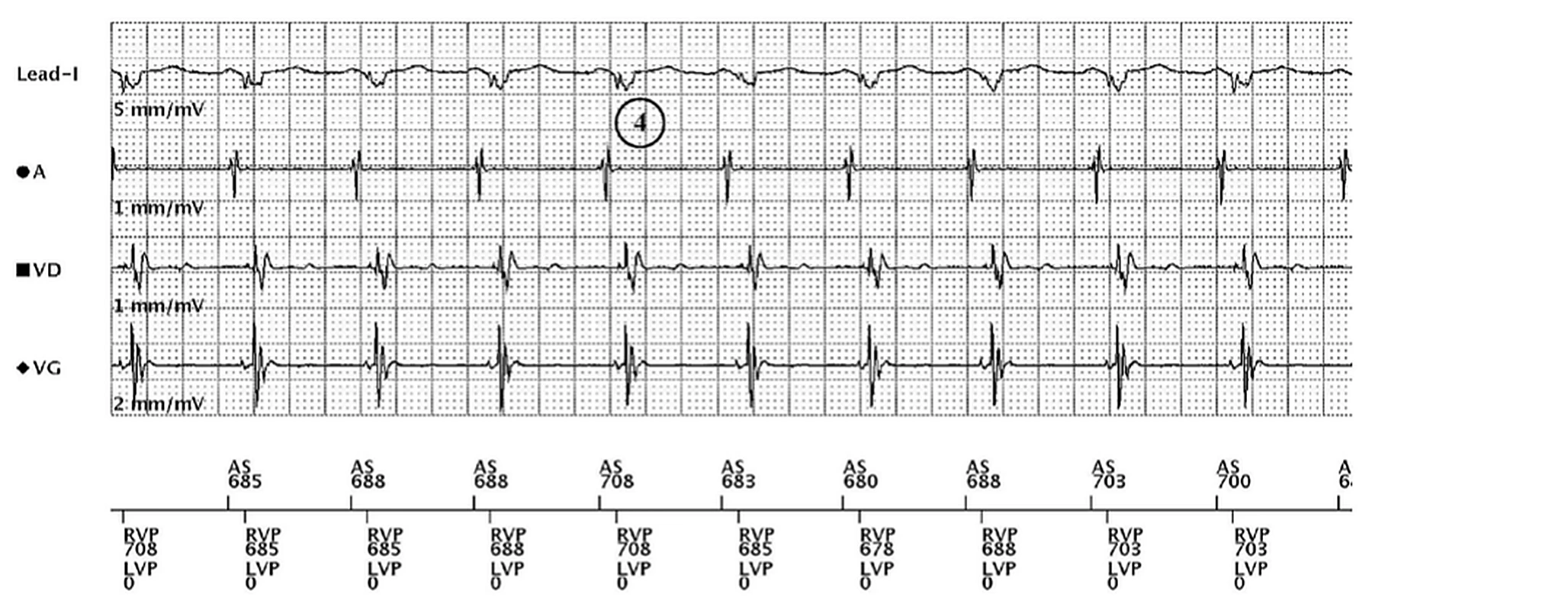
- LV output reprogrammed to 3.5 V/0.4 ms, same pacing configuration: the BiV capture QRS on every beat (negative on Lead I; LV EGM now at 2 mm/mV instead of 0.5). So the first tracing, 100% BiV pacing on the counters, was RV capture only
Points to remember
- The counters count paced beats, not captured ones: an LV output set at the LV threshold gives lost or intermittent LV capture that they do not show
- In a non-responder, look at the paced QRS (on an ECG, or Lead I on the programmer) and measure the LV threshold; a paced QRS that changes from beat to beat points to intermittent LV capture
- Here the chest X-ray showed no visible dislodgement; the LV threshold may have risen from a micro-dislodgement or from local changes in the myocardium
- What to do: measure the LV threshold in each pacing configuration of the lead, keep the one with the lowest threshold without phrenic nerve stimulation, and leave a safety margin above it; here 3.5 V restored BiV capture on every beat
Summary
- The LV threshold has risen to the programmed LV output (2.5 V): LV capture is lost or intermittent while the counters show 100% BiV pacing; at 3.5 V every beat is BiV captured
Clinical pearl
100% BiV pacing on the counters is not 100% BiV capture: in a non-responder, look at the paced QRS and measure the LV threshold
Practice out loud
Just describe the tracing. What do you think is going on? Describing the numbers can help.
00:00
0%
—
From Boston Scientific CRT — clinical cases by M. Strik. Published by Cardiocases. Every numbered marker on a recording is explained in the list beneath it; tap a marker on the trace or an entry in the list.