
Chapter 5 · Bonus · Case 2
Tachycardia while dancing: what the CRT-P and the S-ICD saw
Patient and episode
Patient
- Patient in their mid-forties with dilated cardiomyopathy, on the heart transplant list: VISIONIST X4 CRT-P implanted 6 years earlier, and an EMBLEM MRI S-ICD added 2 months earlier (shock zone 230 bpm, conditional shock zone 200 bpm, SMART Pass on, post-shock pacing off)
- One evening, while dancing: the CRT-P stores episode V-47 “SVT (V<=A)”, A 196 / V 198 bpm, 1 min 48 s; the S-ICD stores episode 001, treated, 2 shocks, shock impedance 133 Ω
The recording
Tap a number on the trace, or an entry in the list below
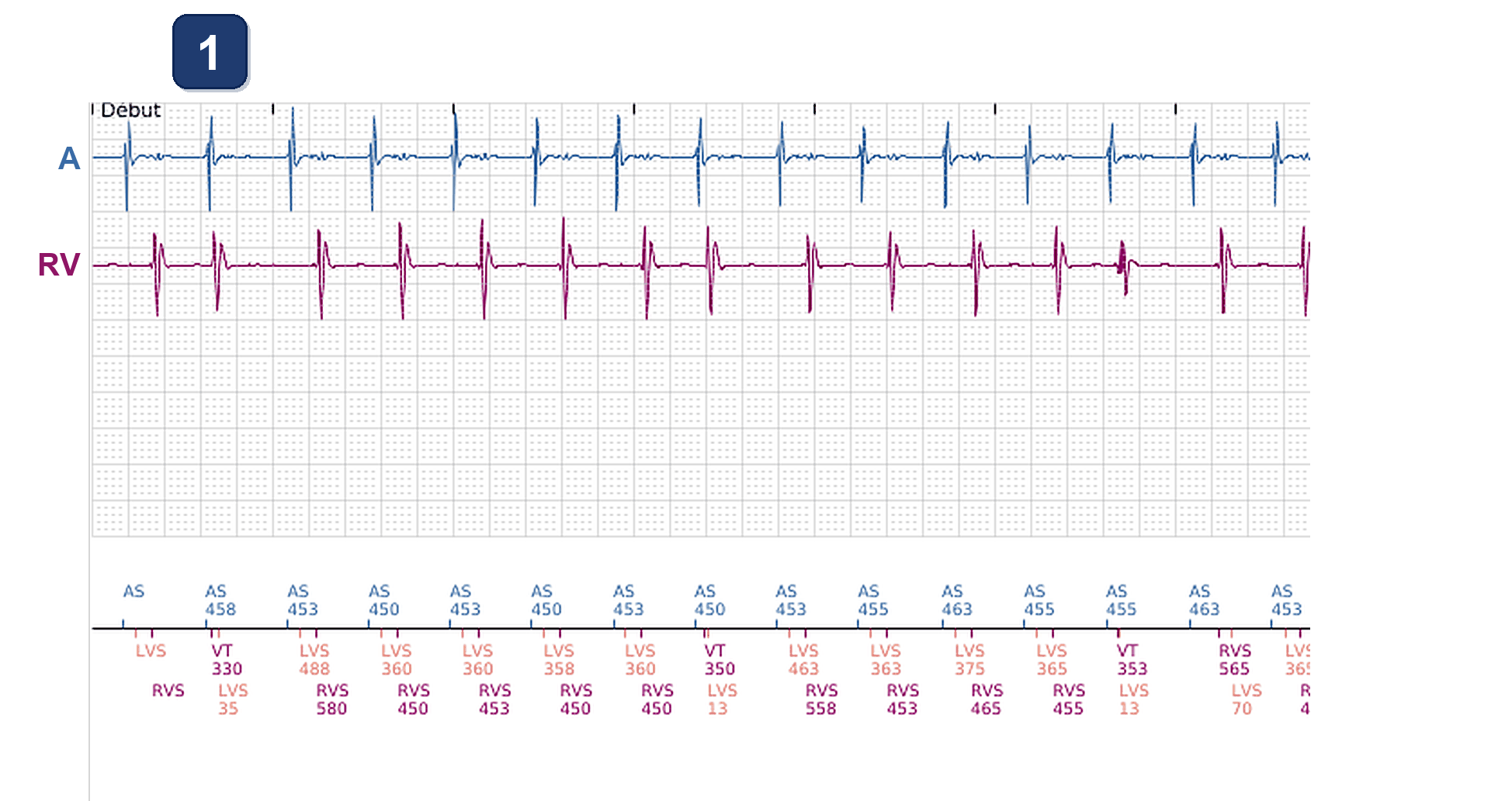
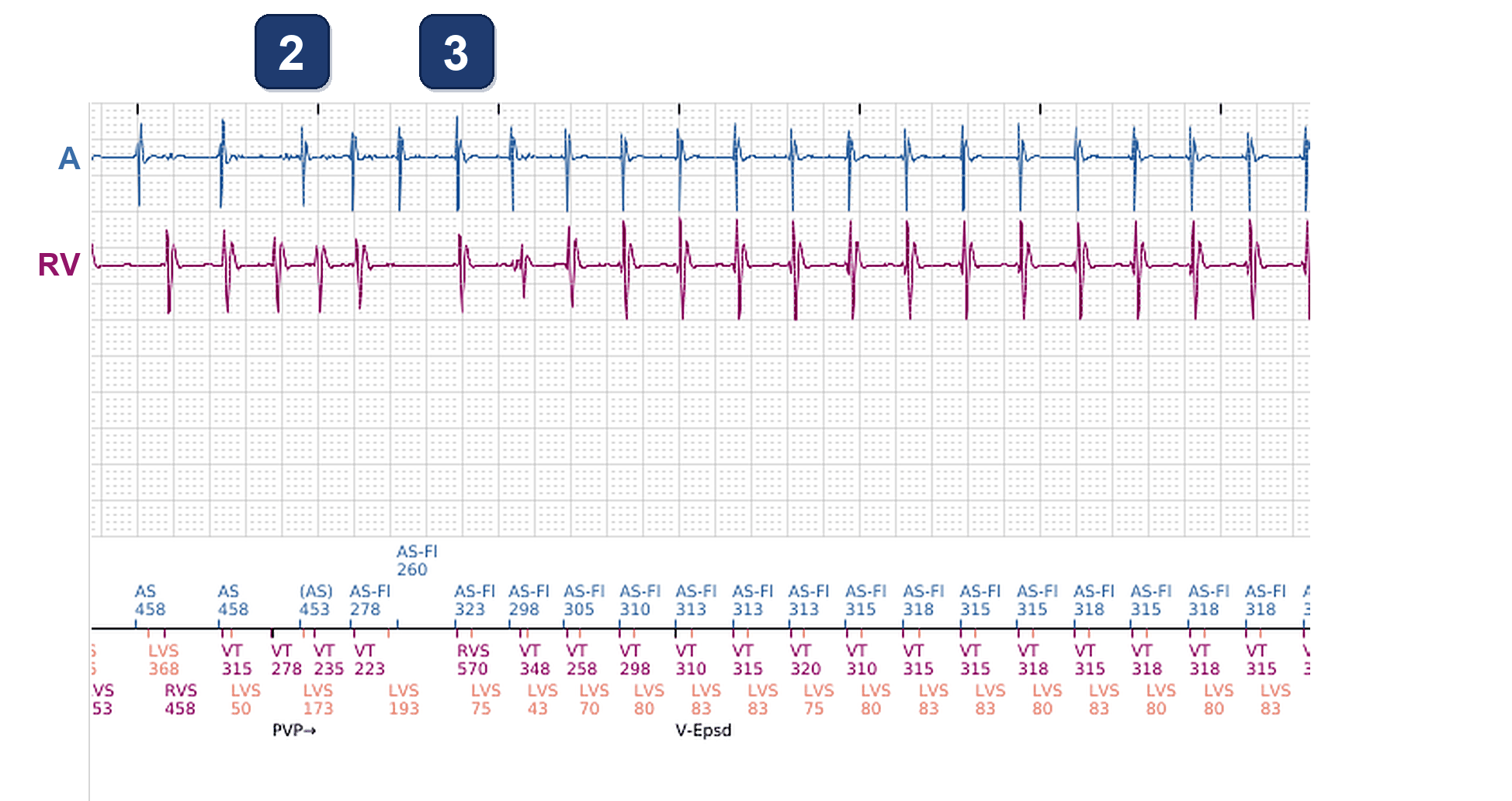
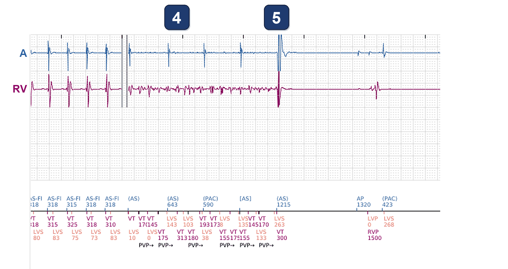
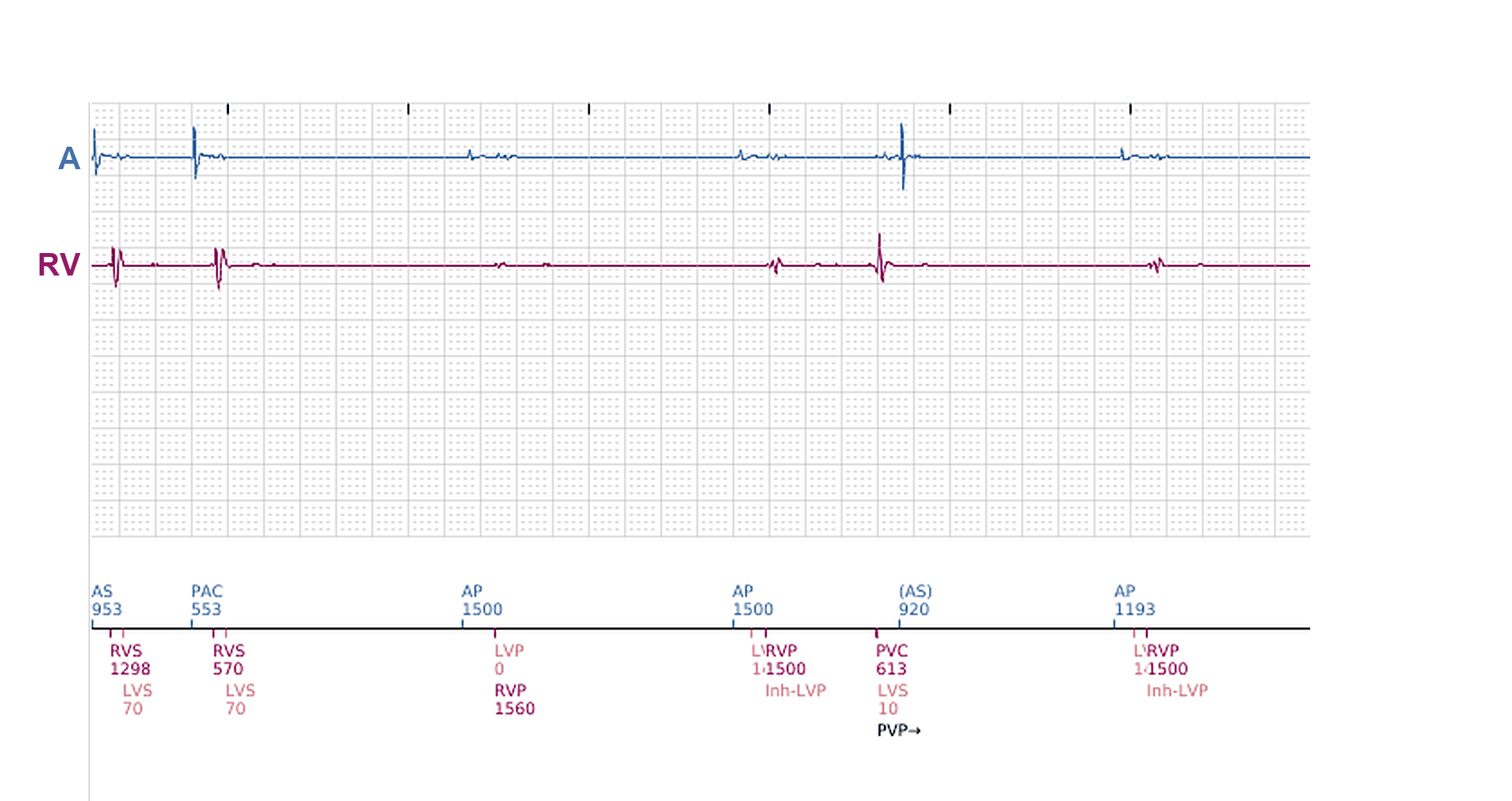
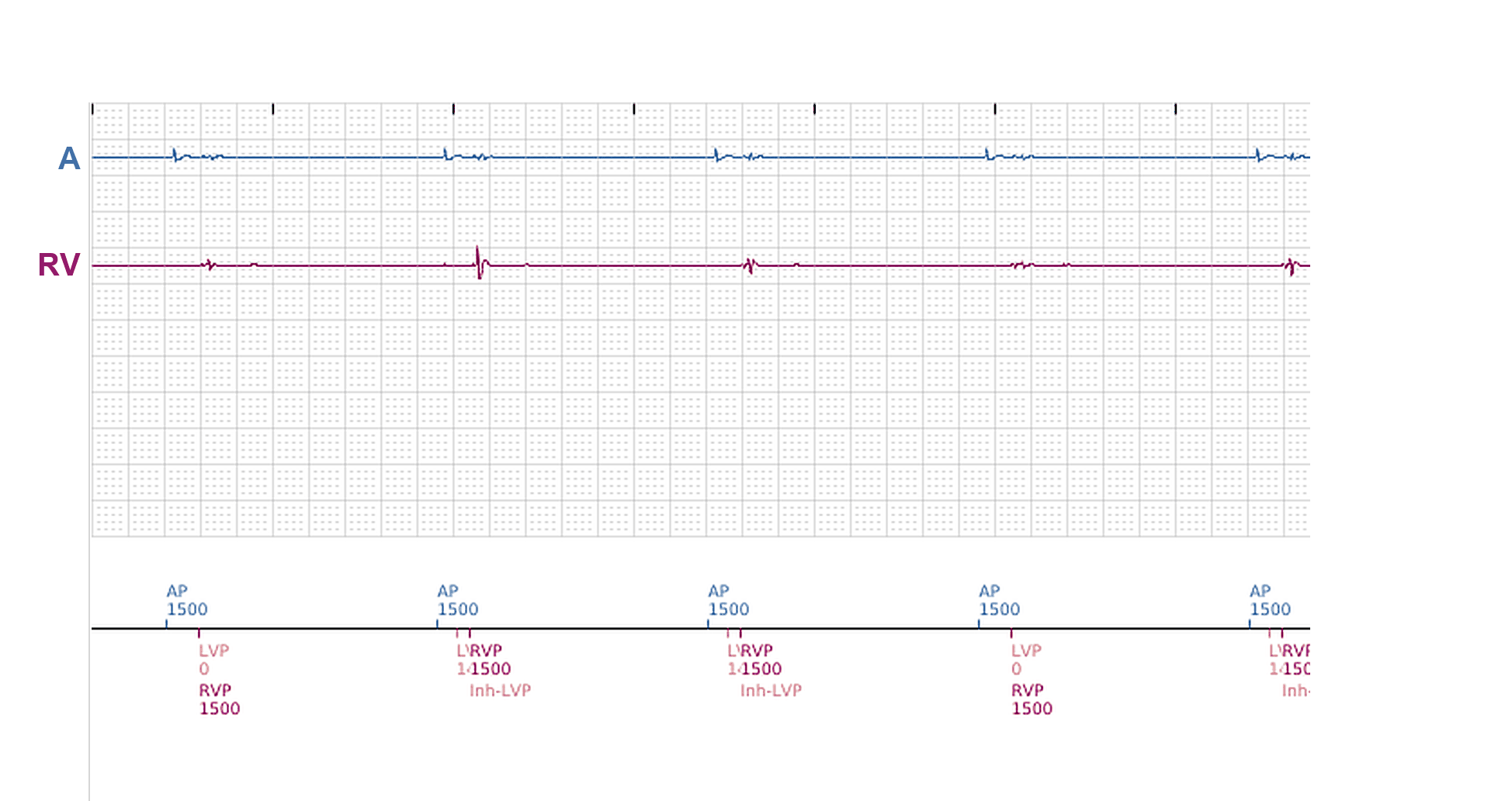
- CRT-P, atrial and RV channels, start of its stored episode: sinus tachycardia while dancing, about 130 bpm. Each atrial event is conducted: the LV is sensed first and the RV about 90 ms later, before the device paces: no BiV pacing. A few isolated PVCs (VT markers)
- The trap: a run of PVCs (VT markers), the RV sensed first and faster than the atrium, which keeps its sinus rhythm. The run is short: it is not the tachycardia, but it triggers it
- After a pause, a regular 1:1 tachycardia at about 190 bpm. At its onset the atrium leads: the atrial cycle changes first and the ventricle follows, the A driving the V, as in an atrial tachycardia conducted 1:1. The atrial events are marked AS-Fl (fast, not tracked); V-Epsd: the CRT-P opens the episode it calls “SVT (V<=A)”, and here the label is right
- Later, after a gap (‖) in the stored EGM: VF, small irregular RV signals (VT markers), while the atrium goes on at its own slower rate
- Shock artefact on both channels: the second S-ICD shock ends the VF. After a pause of about 1.3 s the CRT-P paces the atrium and both ventricles at the lower rate (AP, then LVP and RVP, 40 bpm)
The S-ICD, the same evening
- The S-ICD added two months earlier stored the same event (its clock runs one hour ahead of the CRT-P’s): its S-ECG, from the 1:1 tachycardia to the end
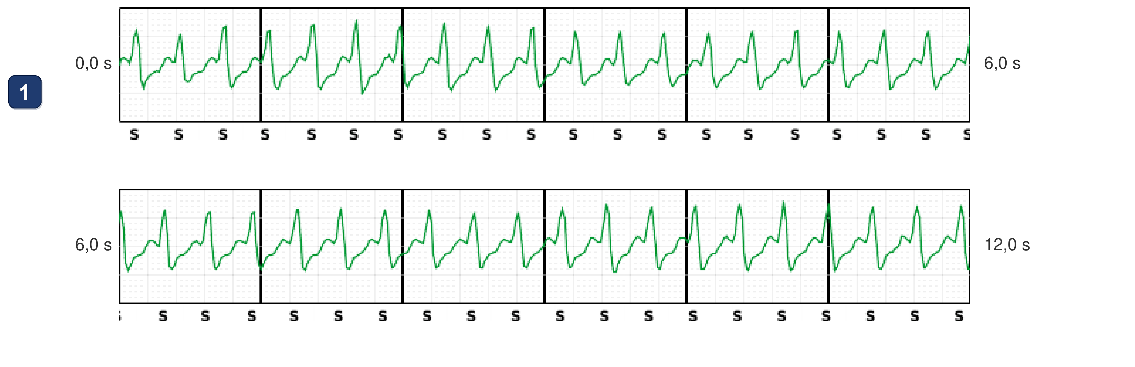
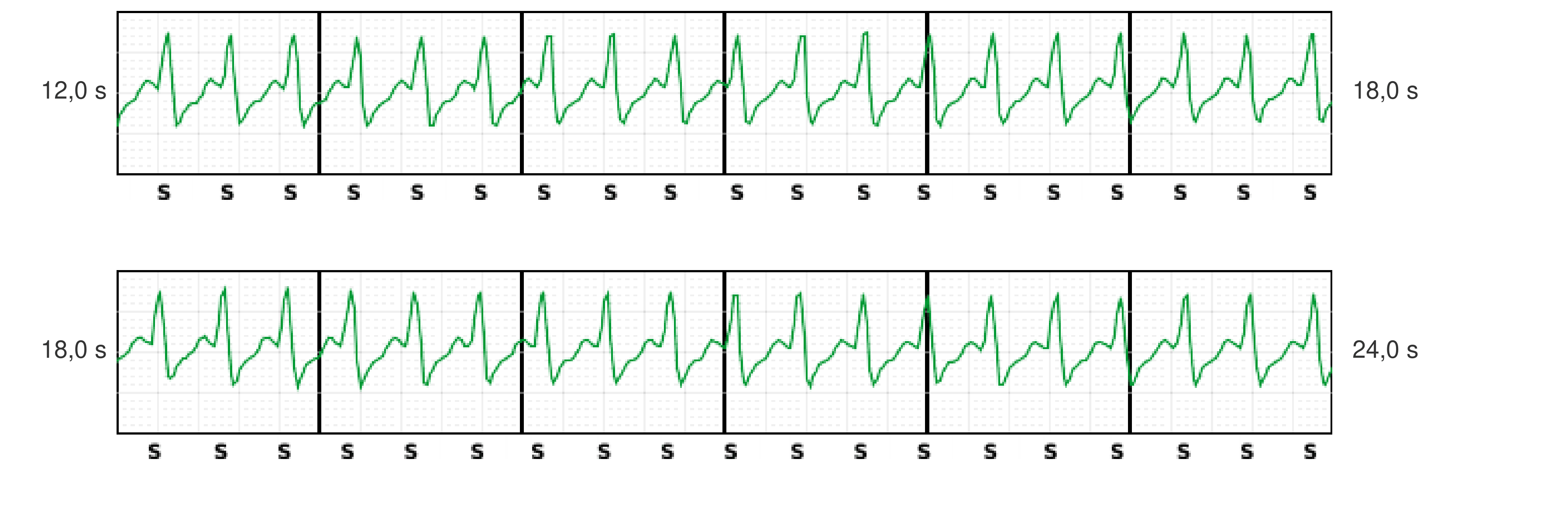
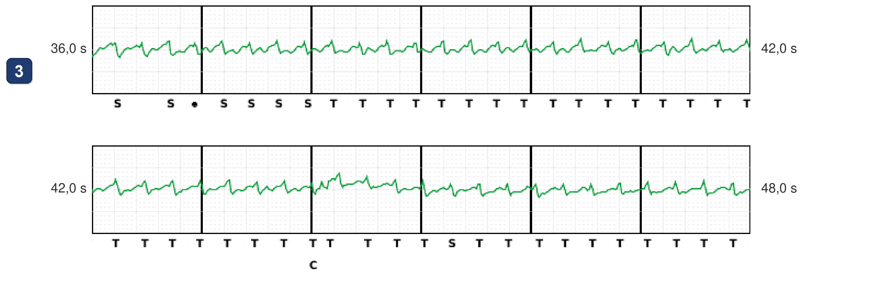
- S-ICD, the 1:1 tachycardia at about 190 bpm: regular complexes, all marked S (sensed), with the same morphology as the patient’s sinus beats: a supraventricular rhythm. Below the conditional shock zone (200 bpm): no treatment
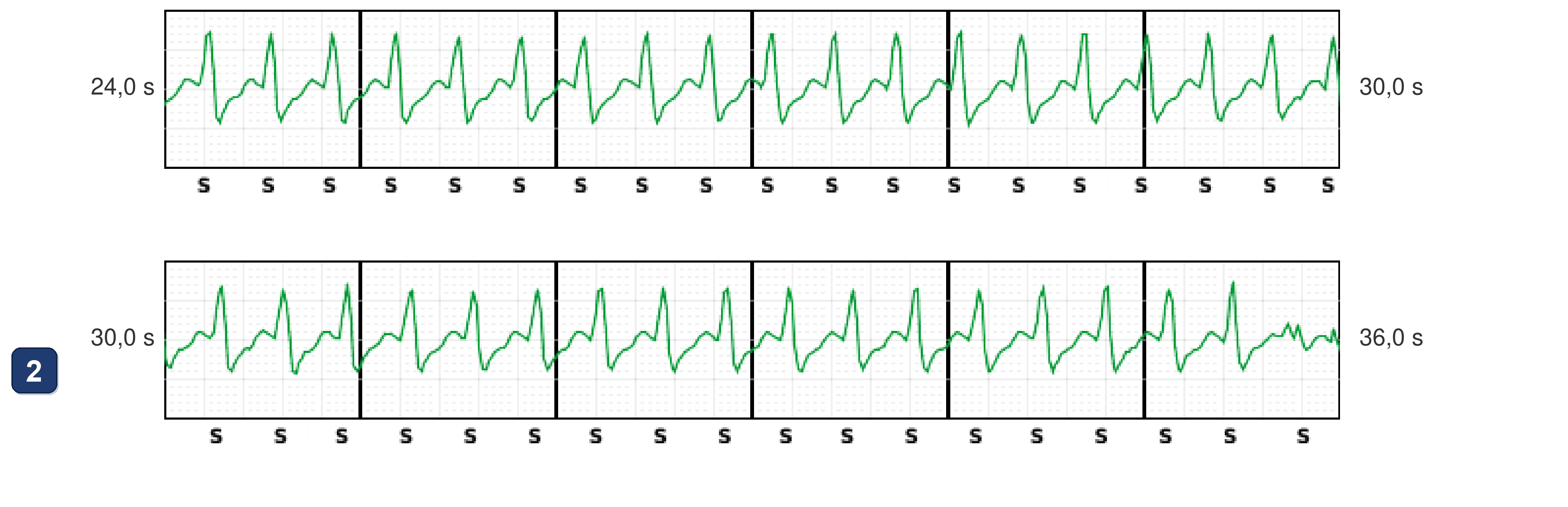
- At about 35.5 s the complexes change: smaller, different and faster (about 240 bpm): the AT has degenerated into a VT
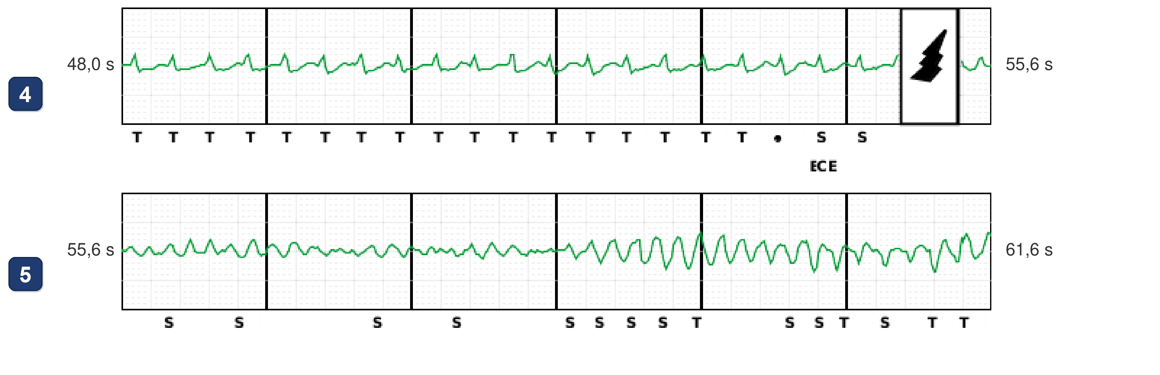
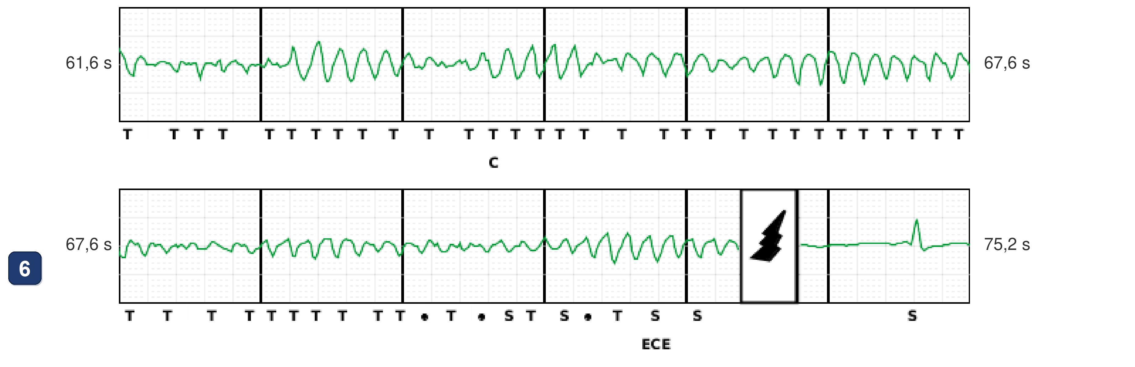
- The VT is in the shock zone (above 230 bpm): beats marked T (tachycardia detection), then the charge starts (C)
- End of charge (E) and first shock at about 54 s
- After the shock the rhythm turns into VF
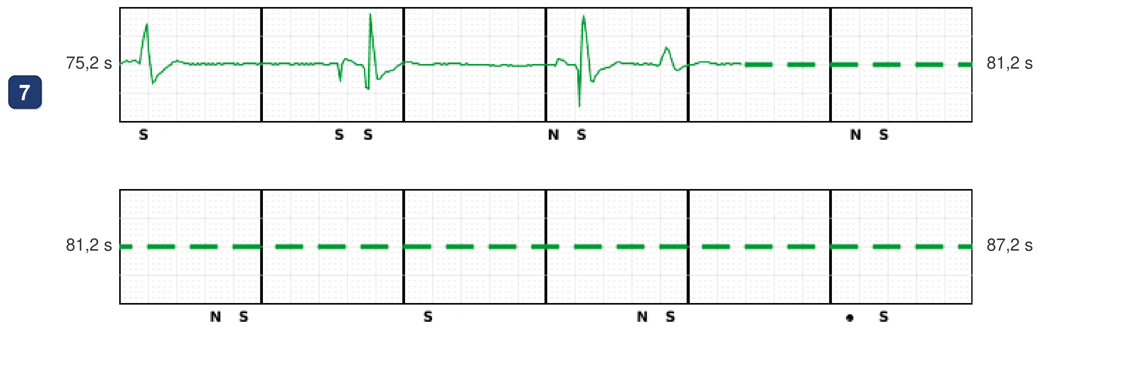
- Second charge (C) and second shock at about 73 s, which ends the VF
- After the second shock: a pause, then slow beats. The S-ECG is no longer stored, but its markers (S) go on at about 120 bpm (report pages 3-4): a sinus tachycardia
Points to remember
- The trap is the run of PVCs: it looks like the start of a VT, but it only triggers the regular tachycardia that follows. Judge that tachycardia by its own onset: here the atrium leads (the A driving the V), as in an atrial tachycardia conducted 1:1
- The S-ICD helps: during the 1:1 tachycardia its complexes keep the sinus morphology, a supraventricular rhythm, which it rightly left untreated below its conditional zone (200 bpm)
- The AT then degenerated into a VT, with smaller, faster complexes (about 240 bpm): the S-ICD detected it in the shock zone and shocked it; VF followed and was shocked again, and the rhythm came back as a sinus tachycardia
- With two devices, line the recordings up by what they show, not by their clocks: here the two clocks were one hour apart for the same event. The CRT-P paced after the shocks (the S-ICD’s post-shock pacing was off)
Summary
- During a sinus tachycardia while dancing, a run of PVCs triggers a 1:1 SVT, probably an atrial tachycardia: the atrium leads at its onset, and the S-ICD complexes keep their sinus morphology. The AT degenerates into a VT, which the S-ICD shocks; VF follows and is shocked again, and a sinus tachycardia returns
Clinical pearl
Sometimes devices can live together: for example a leadless pacemaker as a backup for a malfunctioning pacemaker, an S-ICD on top of a CRT because of venous access problems, or a CRT on top of an S-ICD when LBBB or AV block develops
Practice out loud
Just describe the tracing. What do you think is going on? Describing the numbers can help.
00:00
0%
—
From Boston Scientific CRT — clinical cases by M. Strik. Published by Cardiocases. Every numbered marker on a recording is explained in the list beneath it; tap a marker on the trace or an entry in the list.