Erroneous diagnosis of VT due to T-wave oversensing
Tracing
Manufacturer Medtronic
Device ILR
Field Reveal DX / Reveal XT
N° 10
Patient
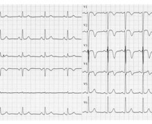
A 54-year old man suffering from ischemic cardiomyopathy with a left ventricular ejection fraction of 56% presented after 2 syncopal episodes. After undergoing negative electrophysiological studies and programmed ventricular stimulation, the patient underwent implantation of a Reveal DX.
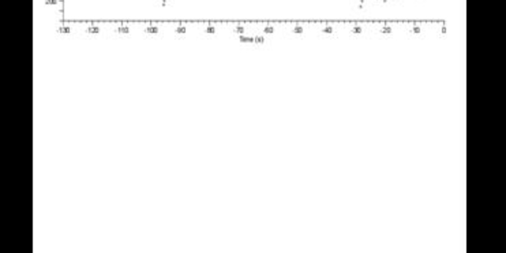
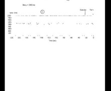
Graph and trace
In absence of recurrent syncope, the implantable Holter recorded 2 FVT and 1 VT episode.
- sinus rhythm;
- railroad-track appearance probably indicative of a supernumerary cardiac signal; the actual ECG recording corresponds to an FVT episode;
- no oversensing in presence of high-amplitude ventricular electrograms;
- oversensing of the T-wave when the R-wave amplitude decreased; whereas the T-wave amplitude remained fixed throughout the recording, the R-wave amplitude varied prominently; the railroad-track appearance of the graph is explained by the alternating incidence of the 2 signals (short RT, long TR);
- a FVT episode was detected.
Other articles that may be of interest to you
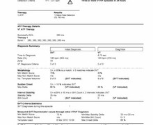

This episode displays characteristics often observed in presence of T-wave oversensing:
The programmable settings that can prevent this oversensing are a) the refractory period, b) the duration of threshold stability before its decrease which, in these patients, must be increased and 3) the sensitivity.