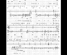
lead fracture and aystole

Tracing
N° 27
Manufacturer Medtronic Device ICD Field Oversensing
Patient

Patient implanted with a triple-chamber ICD (Viva XT CRT-D) for primary dilated cardiomyopathy and hospitalized for syncope; this tracing illustrates the risk of asystole in conjunction with an oversensing caused by lead dysfunction.

Graph and trace

The graph shows initially a large variability of the ventricular intervals with very short intervals followed by longer intervals; stabilization of a very fast ventricular rhythm explaining the sensing of an unsupported episode of VT.

  1. the EGM initially reveals the oversensing of isolated short ventricular intervals related to low amplitude potentials; a right ventricular pacing occurs when a signal is oversensed during the post-atrial pacing window explaining the very short intervals;
  2. the oversensing persists for 2 to 3 seconds explaining the occurrence of a ventricular pause.
Comments

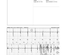
This patient presented an oversensing of pectoral myopotentials which could be reproduced by counter maneuvers of the arm ipsilateral to the pulse generator. In this patient with altered atrioventricular conduction, oversensing induced ventricular pauses of variable duration, responsible for repeated lipothymias/syncopes. Two types of myopotentials can be oversensed by an ICD:

  1. diaphragmatic myopotentials: the use of a high auto-adjusting sensitivity algorithm allows optimizing the quality of the sensing of low-voltage VF signals, but also increases the risk of myopotential oversensing at the end of diastole when sensitivity reaches its maximum. Diaphragmatic myopotential oversensing is rare but has been increasingly observed in patients implanted with an integrated bipolar lead positioned at the apex of the right ventricle. Permanent ventricular pacing is associated with an increased risk of oversensing of these myopotentials given that, after pacing, the time spent at maximum sensitivity is prolonged especially at slow heart rates. An integrated bipolar lead facilitates this phenomenon due to a wider sensing antenna. Diaphragmatic myopotentials correspond to low-amplitude, high frequency signals most often detected exclusively on the near-field channel (absent on the far-field channel). The two main characteristics of this type of signal are that their amplitude varies with the respiratory cycle and that they can be reproduced by specific provocation maneuvers (deep inspiration, Vasalva, forced cough). Oversensing occurs initially at the end of diastole when sensitivity is maximal. Sensing of the true R-wave (of high amplitude) modifies the level of sensitivity and interrupts, at least temporarily, the oversensing of these small signals, which explains why prolonged oversensing only occurs in dependent patients (absence of spontaneous R-wave, sensitivity level permanently at maximum). Oversensing of diaphragmatic myopotentials may be corrected by reducing the sensitivity level, with necessary verification of the reliable sensing of VF signals. In paced patients, an increase in the minimal pacing rate may also have a favorable effect. In some cases, it is necessary to implant a new defibrillation (DF4 system) or pacing (DF1 system) lead remotely from the diaphragm (septum or infundibulum).
  2. pectoral myopotentials: in an ICD, the can being positioned in the pocket in the vicinity of pectoral muscles (which are not part of the sensing circuit), the pectoral myopotentials should not generate oversensing. The amplitude of these myopotentials is greater when recorded at the level of the far-field channel which also includes the can (sensing between the right ventricular coil and the can). On the other hand, if there is an insulation break (typically an erosion leading to a current leak) at the level of the pocket portion of the lead (friction between the can and the lead), then the near-field channel may oversense the pectoral myopotentials, which may lead to inhibition of pacing and/or the occurrence of inappropriate therapies.

Analysis of the EGMs in this setting reveals the presence of very fast (high-frequency) non-physiological signals. Oversensing can be replicated by isometric movements of the arm ipsilateral to the can or by manipulation of the lead in the pocket. When there is suspicion of pectoral myopotential oversensing, a chest X-ray should be performed along with a thorough verification of the device (impedance values, pacing and sensing thresholds). The presence of an abnormally low impedance value (pacing and/or defibrillation) or a sudden decrease in this value is suggestive of an insulation defect. In very rare cases, oversensing of myopotentials can be observed when a DF1 system has been implanted with inversion of the pins in the connector.

Other articles that may be of interest to you