AV and VV delays optimization

Basic concepts

Biventricular resynchronization offers considerable clinical benefits, including reverse remodeling with decrease in cardiac volumes, and a decrease in morbidity and mortality in patients suffering from heart failure with wide QRS complex on the surface electrocardiogram. The main limitation of this therapy is a high percentage of non-responders to the therapy. Various approaches have been proposed to lower that percentage. Once the device has been implanted, the quality of its programming is an important determinant of the quality of the response. CRT aims at changing the sequence of activation in presence of electrical conduction disorders by adjusting the activation delays among the right atrial, RV and LV leads. Two settings can be programmed in this context: 1) the AV delay that determines the activation timing between right atrium and right ventricle, with independent programming of a sensed AV delay, after sensing of a spontaneous atrial event (AS cycle-BV) and a paced AV delay following a paced atrial event (AP-cycle BV). A variable AV delay that decreases in parallel with an increase in heart rate can be programmed; 2) The VV delay is the interval between right and left ventricular activation; simultaneous activation (VV delay = 0), right pre-activation (RV à LV, X ms) or a left pre-activation (LV à RV, X ms) are programmable; a VV delay that varies between rest and exercise cannot be programmed. Short-term hemodynamic studies have found a considerable gain achievable by the optimization of the AV delay, the VV delay or both. The long-term confirmation of this benefit with respect to cardiovascular endpoints (heart failure hospitalization, death or need for heart transplantation) is still lacking.

AV delay optimization

The atrial contraction contributes 20-30% of the cardiac output at rest in patients suffering from heart failure with systolic dysfunction, a contribution that increases with exercise. Patients suffering from heart failure and electrical conduction disorders often present with AV dyssynchrony, shortening of the filling time, merging of the E and A waves and diastolic mitral regurgitation. In patients, undergoing CRT, the programming of a short AV delay advances the E wave, separates it from the A wave and lengthens the filling time. The AV delay should not be excessively short, since it amputates the A wave by closing the mitral valve. Despite modest clinical evidence, it is recommended to adjust the AV delay after implantation of a CRT device.

The inter-individual variations in intra-atrial conduction and intra-ventricular disorders are wide, causing wide variations in optimal AV delay, which theoretically justify individual programming of each device. The separately programmable sensed and paced AV delays must also be optimized independently. The AV delay is usually optimized at rest, in the supine position, during fixed heart rate, conditions that differ considerably from those of everyday life. Unlike in patients with healthy hearts, in whom the optimal AV delay shortens as the heart rate increases during exercise, the response of CRT devices to stress is not predictable since, the optimal AV delay is longer in some patients during exercise than at rest, whereas in others it is shorter. While the systematic use of an automatic AV delay reduction algorithm probably ensures a reliable capture during exercise, it is not necessarily associated with an additional hemodynamic benefit. Therefore its programming should be individualized. BiV resynchronization promotes reverse remodeling and progressively decreases the end-systolic and end-diastolic volumes over time. Therefore, the AV delay must be periodically re-optimized.

An optimal AV delay promotes a maximum contribution of the left atrial contraction to LV filling, lengthens the filling time, increases the cardiac output and minimizes mitral regurgitation. If the length of the AV delay is excessive, the atria contract too soon in diastole, limiting their contribution to ventricular filling. The atrial contraction is superimposed over early diastole, echocardiographically apparent as a fusion of E and A waves, a short filling time and persistent diastolic mitral regurgitation. If the length of the AV delay is insufficient, the ventricles contract too early, causing premature closure of the mitral valve, interrupting on-going filling and limiting the atrial contribution to ventricular filling. Echocardiography shows a premature E wave, a long filling time, split E and A waves and an A wave truncated by the mitral valve closure. The decrease in end-diastolic pressure and preload decreases the peak dP/dt and the cardiac output.

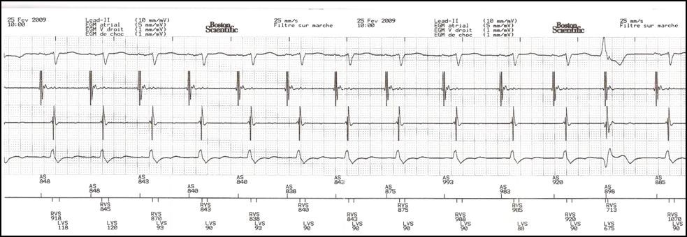
Before optimizing the AV delay, it is useful to keep in mind that, in presence of complete or high-grade AV block or a markedly prolonged PR interval, changing the AV delay has no direct effect on the amount of ventricular capture and fusion. In contrast, in presence of preserved AV conduction, prolonging the AV delay causes progressive fusion of ventricular capture with spontaneous activation. The AV delay must be adjusted under electrocardiographic guidance, remembering that, in absence of complete AV block, which is the case in the majority of patients, that adjustment varies the delay between atrial and ventricular systole, while also directly interfering with the ventricular activation sequence and the amount of ventricular fusion. This can be overcome by systematically programming a short AV delay, between 90 and 120 ms after a sensed P-wave, and between 130 and 150 ms after a paced atrial event.

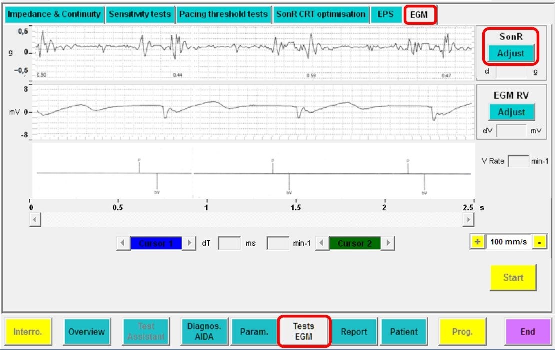
Progressive adjustment of the AV delay in a CRT system recipient with preserved AV conduction. Fusion gradually increases as the AV delay lengthens.

Various means of AV delay optimization have been proposed:

- Echocardiography

A) The Ritter method, which has not been validated in a sample of patients presenting with heart failure; B) search for a maximum aortic or mitral velocity-time integral; C) search for maximum dP/dt; and D) the iterative method. The latter method, widely used in clinical practice, analyses the trans-mitral flow to obtain the longest ventricular filling time without amputation of the A wave.

- Other methods

Cardiac contractility or output can be estimated by examining the pulse wave, blood pressure, maximum dP/dt, electrocardiographic morphology, and others. The applicability of these estimates in daily practice is often limited.

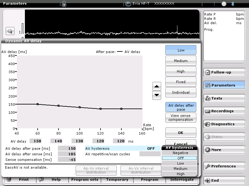
- Embedded automatic optimization algorithm

If the AV delay must be optimized repeatedly and under various conditions of preload, it should ideally be accomplished by the pacemaker. Different algorithms developed by the different companies will be described at the end of this chapter.

VV delay optimization

Considerable ventricular mechanical dyssynchrony may persist after device implantation in non-responders to CRT. Adjustments of the VV delay result in sequential BiV pacing and directly modify the sequence of ventricular activation, which may alleviate the persistent dyssynchrony in non-responders. This setting may be theoretically advantageous in presence of a suboptimal position of the LV lead, or of latency and a prolonged conduction time to the site of stimulation. While the optimization of the VV delay has been associated with significant short-term hemodynamic benefits, the long-term clinical relevance of this programming remains debated and unconfirmed by clinical studies. The remodeling process probably directly influences the VV delay optimization, such that it must be re-optimized over time under various conditions of preload.

The same methods can be used to optimize the AV and VV delays. Echocardiography is often used in clinical practice. Aortic velocity-time integral, which reflects the cardiac output, maximum dP/dt, which reflects cardiac contractility, or measurement of the degree of ventricular asynchrony are also used. In light of the practical difficulties of VV optimization, its repeated automatic adjustment by the device is a promising development. However, its clinical relevance remains to be confirmed.

Influence of the VV delay on the ventricular electrical activation

 Despite the evident difference in electrocardiographic morphology among the various configurations, which one is likely to be associated with an optimal clinical response is not clear.

LV alone versus BiV stimulation

The superiority of BiV over LV stimulation has not been confirmed, though short-term hemodynamic studies have consistently found a significant benefit conferred by isolated LV stimulation. Similarly, clinical studies have observed a nearly identical benefit conferred by isolated LV stimulation on NYHA functional class, exercise capacity and ventricular remodeling compared with BiV stimulation. However, all large studies demonstrating the benefits conferred by CRT have used BiV instead of LV stimulation alone. With that latter configuration, the resynchronization of the two ventricles is achieved by the fusion between the stimulated left ventricle and the spontaneous RV activation. The optimal short-term hemodynamic benefit seems to be achievable with an optimal degree of fusion, which is difficult to quantitate and preserve during exercise, due to changes in the heart rate and PR interval.

Isolated LV stimulation is an attractive option, particularly when the implanted device is a CRT-P. This can be accomplished with a dual chamber pacemaker without implanting an RV lead, which lowers the cost of the system and lowers the risk of complications. However, in pacemaker-dependent patients presenting with AV block, the implantation of a single LV lead may be risky, given the high incidence of LV lead dislodgement, rise in capture threshold, or both. In recipients of CRT-D, the implantation of a RV lead is indispensible. However, programming of the device in a “LV stimulation only” configuration eliminates the battery consumption associated with RV stimulation.

Specificities

As mentioned earlier, a repetitive optimization of the stimulation configuration over time should ideally be performed automatically by the device. This optimization should be effortless for the caregiver, without burdening the medical services such as echocardiography or electrophysiology laboratories and, as in the case of most measurements made by the device, should be reproducible. Several algorithms have been developed to satisfy these needs.

Biotronik

This manufacturer has not developed a specific AV and VV delay optimization algorithm. On the other hand, a negative hysteresis of the AV delay can be programmed in order to trigger BiV stimulation. In a pacemaker non-dependent patient, the programmed AV delay can be shortened if it is longer than the spontaneous AV interval.

Boston Scientific

The SmartDelay® function enables the optimization of the sensed and paced AV delays, as well as a choice of LV versus BiV stimulation, using abacuses recorded in the device. This algorithm is based on the measurements of, respectively, the right and left AV intervals during spontaneous and paced atrial activity, taking in consideration a programmed V-V delay. This optimization procedure can be performed within 2.5 min with the programmer during ambulatory visits, though is not automatically repeated by the device during follow-up.

Measurement of right and left AV delays during spontaneous atrial activity

Atrial pacing at 80 bpm and measurement of right and left AV intervals during paced atrial activity

Once these measurements have been made, the algorithm proceeds with the setting of the sensed and paced AV delays and choice of the stimulated chamber(s).

Medtronic

Operating principles of the AdaptivCRT® algorithm

The AdaptivCRT algorithm is activated by selecting 1) the “Adaptive BiV” setting, whereby the device optimizes automatically the AV and VV delays, 2) the “Adaptive BiV and LV” settings, whereby it chooses between LV stimulation alone with optimization of the AV delay versus regular BiV stimulation with optimization of the AV and VV delays, or 3) “Nonadaptive CRT”, in which case the algorithm is deactivated. The range of sensed AV delays of the AdaptivCRT function is limited to between 80 and 140 ms, while the paced AV delays range between 100 and 180 ms. The timings of the VV delays (left or right pre-excitation) range between 0 ms and 40 ms.

The AdaptivCRT operating function relies on the regular evaluation of 1) the AV conduction time, which corresponds to the delay between the EGM recorded by the right atrial lead and that recorded by the RV lead, 2) the P wave duration, which corresponds to the delay between the atrial EGM recorded on the bipolar channel of the right atrial lead and the end of the atrial EGM recorded by the high-voltage channel, and 3) the width of the QRS complex, which corresponds to the delay between the EGM detected by the RV bipole and the end of the EGM recorded on the high-voltage channel. The algorithm measures the patient’s spontaneous AV conduction every minute to determine whether it is normal or prolonged. The AV interval is measured by extending the sensed and paced AV delay to 300 ms to enable spontaneous conduction. In absence of >3 spontaneously conducted consecutive ventricular cycles, prolonged AV conduction is diagnosed and the time interval between each measurement of the AV interval doubles, from a minimum of 2 min to a maximum of 16 h. The measurements of P wave and QRS durations are scheduled every 16 h. This interval warrants a sampling at various time of the day. During the measurement, the device switches the EGM 1 channel recording to RV coil (HVA)/SVC coil (HVB), or in absence of SVC coil, to HVA/atrial anode. The delay between the atrial and ventricular EGM, and the width of the P wave and QRS are measured after 5 cycles. The first measurements of P wave and QRS duration are scheduled at 30 min after device implant. Thereafter, the P wave and QRS duration can be measured anytime by programming the AdaptivCRT function.

An AdaptivCRT set to “BiV and LV”, can automatically switch between auto BiV and LV mode. Pure LV mode stimulation is expected if 1) the heart rate is ≤ 100 bpm, 2) the delay between the spontaneous atrial and spontaneous ventricular EGM is ≤ 200 ms, and 3) the delay between the paced atrial and the spontaneous ventricular EGM is ≤ 250 ms. In absence of all these criteria, the patient is stimulated in BiV mode.

Details of the algorithm operating function

The device begins with an examination of spontaneous conduction to determine whether the AV interval is normal or prolonged. A normal atrial-sensed AV interval is defined as <200 ms and a normal atrial-paced interval as <250 ms. In presence of normal AV conduction and a heart rate <100 bpm, the Adaptative LV stimulation mode (LV stimulation only) is applied. The timing of LV stimulation is automatically adjusted based on minute-by-minute measurements of the spontaneous AV interval. If the AV conduction time exceeds 133.3 ms, LV stimulation is delivered at approximately 70% of the spontaneous AV interval. If the AV conduction time is < 133.3 ms, LV stimulation is delivered 40 ms before the spontaneous QRS (calculated AV delay - 40 ms). If the spontaneous AV interval is prolonged, or the heart rate is >100 bpm, or loss of LV capture is confirmed by the LV Capture Management®, the Adaptive BiV mode is activated. 

The AV delay will then be calculated as follows:

  • After a sensed atrial event, the AV delay is set to pace 40 ms after the end of the P wave (measured on the high-voltage channel), ≥ 50 ms before the onset of the spontaneous QRS.
  • After a paced atrial event, the AV delay is set to pace 30 ms after the end of the P wave (measured on the high-voltage channel), ≥50 ms before the onset of the spontaneous QRS during atrial pacing, between the atrial stimulus and the bipolar RV EGM.

During Adaptive BiV stimulation, the optimal VV delay is derived from the QRS duration. If the QRS duration (between the bipolar RV EGM and the end of the ventricular EGM on the high-voltage channel) is between 50 and 150 ms, the left ventricle is pre-excited. If the QRS duration is between 150 and 180 ms, the right ventricle is pre-excited. If the QRS duration is not included between 50 and 180 ms, a 10-ms LV or RV pre-excitation is applied. The AV conduction times and P wave duration are also used to optimize the VV delay. If the AV interval during spontaneous atrial rhythm is longer than the P wave, the VV delay is set at 0 ms.

Abbott

The QuickOpt™ algorithm optimizes the paced and sensed AV delay, and the VV delay based on the P-wave duration, measured from the atrial EGM (AV delays) and on the measurement of the VV delays during spontaneous rhythm, RV stimulation and LV stimulation (VV delay).

Four measurements are made automatically:

- P-wave duration
- VV delay during spontaneous rhythm
- VV delay during RV stimulation
- VV delay during LV stimulation

Optimization of the AV delay

The optimal post sensed AV delay corresponds to the P-wave duration + 30 ms if the P-wave duration is >100 ms, and to the P-wave duration + 60 ms if the P-wave duration is <100 ms. The post stimulation AV delay = the post sensed AV delay + 50 ms.

Measurement of P-wave duration

Optimization of the VV delay

The optimal VV delay is calculated from the formula: ΔV spontaneous + (tLR – tRL) ) / 2

Where spontaneous ΔV = the delay between right and left activation during spontaneous rhythm; tLR  = VV interval during LV stimulation; tRL : interval between RV and LV during RV stimulation


Microport CRM-Sorin

The SonR® micro-accelerometer hemodynamic sensor is enclosed in a sealed capsule at the tip of an atrial pacing lead (IS-1 compatible SonRtip lead).

The sensor measures in g (m/s2) the myocardial micro-accelerations throughout the cardiac cycle; SonR1, one of its main components, is created by the iso-volumic contraction.

The SonR1 component of this signal corresponds to the first heart sound S1. Its variations are correlated to the variations in LV maxdP/dt.

The SonR CRT-D systems digitize and analyze the SonR1 signal originating from the atrial lead. The algorithm contained in these defibrillators automatically optimizes the AV and VV delays on a weekly basis.

The SonR optimization, carried out in the night of Sunday to Monday, is an automatic weekly test of 69 combinations of AV/VV delays. Each combination of AV/VV delays is adjusted in a sequence of 3 adaptive cycles followed by 6 cycles to measure the SonR.

Test of VV delay optimization: Monday at midnight:

Seven BiV configurations are tested (from LV à RV 48 ms to RV à LV 48 ms; in 16 ms steps) using 6 different AV delays, from 30 ms to spontaneous conduction minus 50 ms.
This test includes 42 combinations and 252 SonR measurements.

Test of AV delay optimization at rest: Monday à 01:00h:

After optimization of the VV delay, 11 different AV delays, from 30 ms to spontaneous conduction minus 50 ms are tested. This test includes 11 combinations and 66 SonR measurements.

Test of paced AV delay optimization: Monday at 02:00h:

After optimization of the VV delay, 11 different AV delays, from 30 ms to spontaneous conduction minus 50 ms, are tested. This test includes 11 combinations and 66 SonR measurements.

Test of AV delay optimization during exercise: from Monday at 12:00h:

Optimization of the AV delay during exercise starts as soon as the heart rate has reached a programmed value. After optimization of the VV delay, 5 different AV delays, from 30 ms to spontaneous conduction during exercise minus 50 ms, are tested. This test includes 5 combinations and 30 SonR measurements.

Condition of implementation of AV delays or optimized VV delays:

A new VV delay is implemented only after a ≥ 14% increase in SonR1. If the new optimal VV delay differs from the previous VV delay by > 16 ms, the VV delay is progressively increased in 16 ms steps until this new optimal VV delay has been reached.

The variation in AV delay is systematically limited to 20 ms. The ranges are:

- Sensed AV delay at rest = [60-180 ms]
- Paced AV delay at rest = [92-240 ms]

Manual test

Tests of AV/ VV delays optimization can also be performed during the patient follow-up, including with the LV only configuration.

Memorized data

The average amplitude of the SonR signal and the adaptation of the AV and VV delays optimized weekly are available in the memories of the SonR CRT-D.

The SonR signal is also recorded during episodes of tachyarrhythmia in order to examine the immediate changes in cardiac contractility and evaluate the hemodynamic tolerance of an arrhythmia.

Fall in the amplitude of the SonR signal at the time of non-sustained ventricular tachycardia

The SonR CRT-D can also transmit real-time SonR signals by telemetry.

Real-time inspection of SonR signal